 Login / Register


- Clone
- SMI 52 (See other available formats)
- Regulatory Status
- RUO
- Other Names
- Microtubule-associated protein 2, MAP-2
- Previously
-
Covance Catalog# SMI-52P
- Isotype
- Mouse IgG1, κ
- Ave. Rating
- Submit a Review
- Product Citations
- publications

-

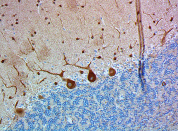
IHC staining of purified anti-MAP2 antibody (clone SMI 52) on formalin-fixed paraffin-embedded mouse brain tissue. Following antigen retrieval using Sodium Citrate H.I.E.R., the tissue was incubated with 0.16 µg/mL of the primary antibody for 60 minutes at room temperature. BioLegend's Ultra-Streptavidin (USA) HRP Detection Kit (Multi-Species, DAB, Cat. No. 929901) was used for detection followed by hematoxylin counterstaining, according to the protocol provided. The image was captured with a 40X objective. Scale bar: 50 µm -

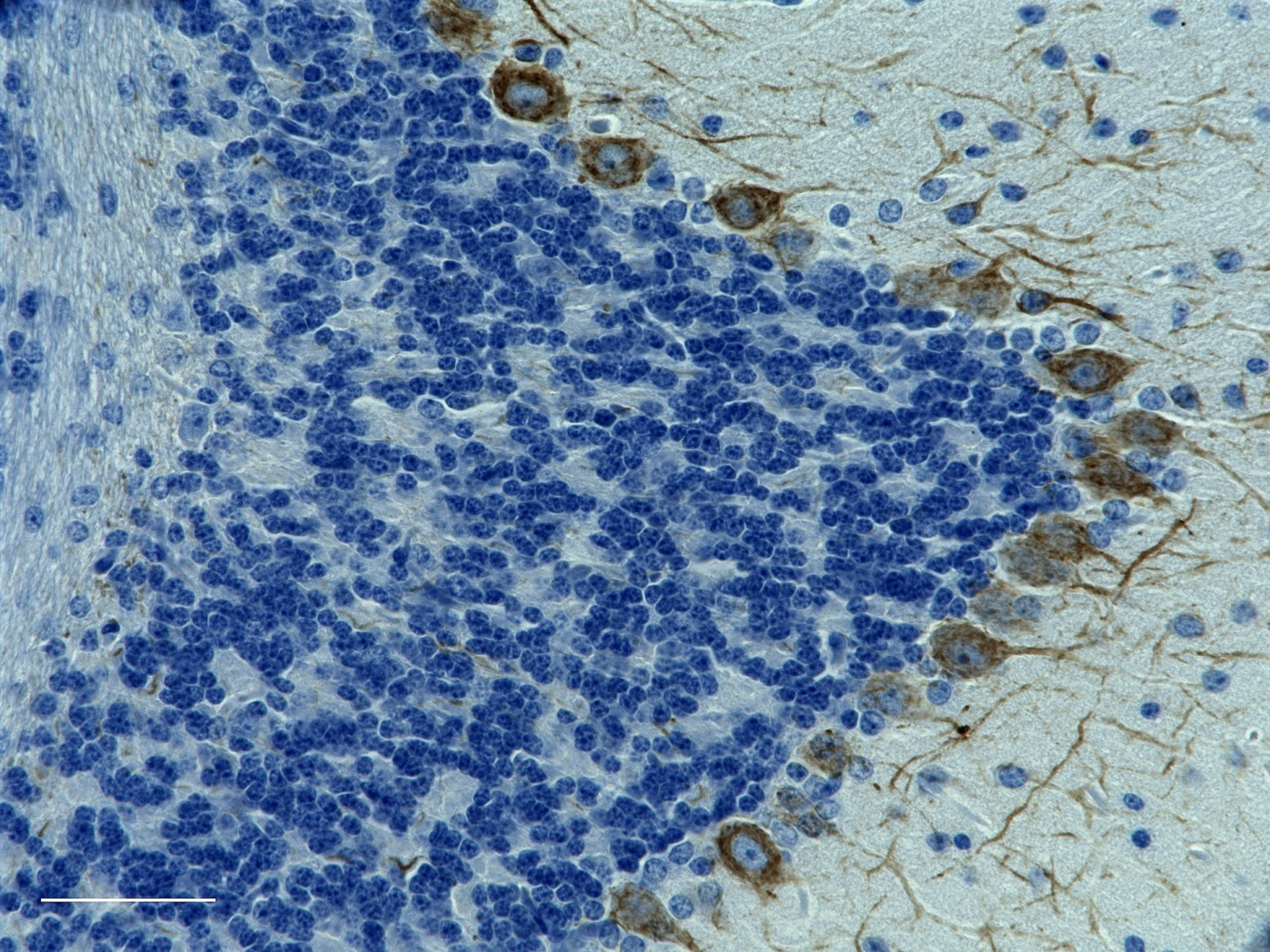
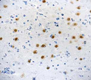
IHC staining of purified anti-MAP2 antibody (clone SMI 52) on formalin-fixed paraffin-embedded rat brain tissue. Following antigen retrieval using Sodium Citrate H.I.E.R., the tissue was incubated with 0.16 µg/mL of the primary antibody for 60 minutes at room temperature. BioLegend's Ultra-Streptavidin (USA) HRP Detection Kit (Multi-Species, DAB, Cat. No. 929901) was used for detection followed by hematoxylin counterstaining, according to the protocol provided. The image was captured with a 40X objective. Scale bar: 50 µm -

IHC staining of purified anti-MAP2 antibody (clone SMI 52) on formalin-fixed paraffin-embedded rat brain tissue. Following antigen retrieval using Sodium Citrate H.I.E.R., the tissue was incubated with 0.16 µg/mL of the primary antibody for 60 minutes at room temperature. BioLegend's Ultra-Streptavidin (USA) HRP Detection Kit (Multi-Species, DAB, Cat. No. 929901) was used for detection followed by hematoxylin counterstaining, according to the protocol provided. The image was captured with a 40X objective. Scale bar: 50 µm
| Cat # | Size | Price | Quantity Check Availability | Save | ||
|---|---|---|---|---|---|---|
| 801810 | 25 µL | 112 CHF | ||||
| 801801 | 100 µL | 275 CHF | ||||
Microtubules are 25nm diameter protein rods found in most kinds of eukaryote cells. They are polymerized from a dimeric subunit made of one α subunit and one β tubulin subunit. Microtubules are associated with a family of proteins called microtubule associated proteins (MAPs), which includes the protein τ (tau) and a group of proteins referred to as MAP1, MAP2, MAP3, MAP4 and MAP5.
Product DetailsProduct Details
- Verified Reactivity
- Mouse, Rat
- Antibody Type
- Monoclonal
- Host Species
- Mouse
- Immunogen
- This monoclonal antibody was raised against Microtubule-Associated Protein 2 (MAP2).
- Formulation
- Phosphate-buffered solution.
- Preparation
- The antibody was purified by affinity chromatography.
- Concentration
- 1 mg/mL
- Storage & Handling
- The antibody solution should be stored undiluted between 2°C and 8°C. Please note the storage condition for this antibody has been changed from -20°C to between 2°C and 8°C. You can also check your vial or your CoA to find the most accurate storage condition for this antibody.
- Application
-
IHC-P - Quality tested
ICC, ELISA, WB - Reported in the literature, not verified in house - Recommended Usage
-
Each lot of this antibody is quality control tested by formalin-fixed paraffin-embedded immunohistochemical staining. For immunohistochemistry, a concentration range of 0.16 - 1.0 µg/mL is suggested. It is recommended that the reagent be titrated for optimal performance for each application.
- Application Notes
-
Additional reported applications (for relevant formats) include: immunocytochemistry5,6, ELISA7, and WB1,5.
SMI 52 reacts with microtubule-associated protein 2 (MAP2). This antibody reacts with most mammalian species and with frog tissue. SMI 52 recognizes neuronal cell bodies and dendrites in tissue sections and cell cultures. In immunoblots of brain cytoskeletal preparations, SMI 52 reacts with MAP2a and b, and also with a doublet at 68 kD that may represent MAP2c. -
Application References
(PubMed link indicates BioLegend citation) -
- Shi Y, et al. 2017. Cell Death Differ. 24(1):167. (WB)
- Shelton MA, et al. 2015. Biol. Psychiatry. 78(6):374. (IHC) PubMed
- Wang X, et al. 2015. PLoS One. 10: 0145441. (IHC) PubMed
- Kazim SF, et al. 2014. Neuro Dis. 71:110. (IHC)
- Cardenas-Aguayo MC, et al. 2013. PLOS ONE. (1):e53596. (ICC, WB) PubMed
- Lo Furno D, et al. 2013. J. Cell. Physiol. 228:2109. (ICC)
- Gensel J, et al. 2009. J Neurosci. 29:3956-3968. (ELISA) PubMed
- Product Citations
-
- RRID
-
AB_2728526 (BioLegend Cat. No. 801810)
AB_2564643 (BioLegend Cat. No. 801801)
Antigen Details
- Structure
- MAP2 is made up of two bands of an apparent molecular weight of approximately 280 kD, referred to as MAP2a and MAP2b. A third lower molecular weight form, usually called MAP2c, corresponds to a pair of protein bands running at ~70 kD on SDS-PAGE gels. All these MAP2 forms are derived from a single gene by alternative transcription, and all share a C-terminal sequence which includes either three or four microtubule binding peptide sequences, which are very similar to those found in the related microtubule binding protein τ (tau).
- Distribution
-
Cellular distribution: Plasma membrane, cytoskeleton, nucleus, and cytosol.
Tissue distribution: Predominantly in the nervous system - Function
- MAP2 encodes a protein that belongs to the microtubule-associated protein family. The proteins of this family are involved in microtubule assembly. The products of these genes are neuron-specific cytoskeletal proteins that are enriched in dendrites, which implicate a role in determining and stabilizing dentritic shape during neuron development.
- Interaction
- MAP2 interacts with microtubules. It has also been reported to interact with KNDC1 via KIND2.
- Ligand/Receptor
- MAP2 isoforms are expressed in neuronal cells and specifically in the perikarya and dendrites of these cells. Antibodies to MAP2 are therefore excellent markers on neuronal cells, their perikarya and neuronal dendrites. In contrast τ (tau) is found predominantly in neuronal axons.
- Cell Type
- Neural Stem Cells
- Biology Area
- Cell Biology, Neuroscience, Neuroscience Cell Markers, Stem Cells
- Molecular Family
- Microtubules
- Antigen References
-
- Zhou Y, et al. 2015, Neuro Oncol. 17(12):1578.
- Loeser H, et al. 2015, Am J Nephrol. 41(3):191.
- Gene ID
- 17756 View all products for this Gene ID 25595 View all products for this Gene ID
- UniProt
- View information about MAP2 on UniProt.org
Related Pages & Pathways
Pathways


Other Formats
View All MAP2 Reagents Request Custom Conjugation| Description | Clone | Applications |
|---|---|---|
| Purified anti-MAP2 | SMI 52 | IHC-P,ICC,ELISA,WB |
| Alexa Fluor® 594 anti-MAP2 | SMI 52 | IHC-P |
| Alexa Fluor® 488 anti-MAP2 | SMI 52 | IHC-P |
| Alexa Fluor® 647 anti-MAP2 | SMI 52 | IHC-P,3D IHC |
Customers Also Purchased
Compare Data Across All Formats
This data display is provided for general comparisons between formats.
Your actual data may vary due to variations in samples, target cells, instruments and their settings, staining conditions, and other factors.
If you need assistance with selecting the best format contact our expert technical support team.
-
Purified anti-MAP2

IHC staining of purified anti-MAP2 antibody (clone SMI 52) o... 
IHC staining of purified anti-MAP2 antibody (clone SMI 52) o... 
IHC staining of purified anti-MAP2 antibody (clone SMI 52) o... -
Alexa Fluor® 594 anti-MAP2

IHC staining of Alexa Fluor® 594 anti-MAP2 antibody (clone S... -
Alexa Fluor® 488 anti-MAP2

IHC staining of Alexa Fluor® 488 anti-MAP2 antibody (clone S... 
IHC staining of Alexa Fluor® 488 anti-MAP2 antibody (clone S... 
IHC staining of Alexa Fluor® 488 anti-MAP2 antibody (clone S... -
Alexa Fluor® 647 anti-MAP2

IHC staining of Alexa Fluor® 647 anti-MAP2 antibody (clone S... 
IHC staining of Alexa Fluor® 647 anti-MAP2 antibody (clone S... Formalin-fixed, 300 micron-thick mouse brain (cerebellum) se...


Follow Us