 Login / Register


- Regulatory Status
- RUO
- Other Names
- 5-(and 6)-Carboxyfluorescein diacetate succinimidyl ester Kit, CFDA SE Kit
- Ave. Rating
- Submit a Review
- Product Citations
- publications
-

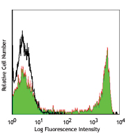
Human peripheral blood mononuclear cells were stained with CFSE Cell Division Tracking Kit, and then stimulated with (filled histogram) or without (open histogram) PHA for 5 days. On day 5, cells were harvested and the CFSE fluorescent staining was analyzed by flow cytometry.
| Cat # | Size | Price | Quantity Check Availability | Save | ||
|---|---|---|---|---|---|---|
| 423801 | 1 kit | £70 | ||||
CFSE Cell Division Tracker Kit is composed of 5 vials, 100 µg per vial of CFSE (formally known as 5-(and 6)-Carboxyfluorescein diacetate succinimidyl ester of CFDA SE), and 500 µl of anhydrous DMSO. CFSE is able to passively diffuse into cells. Inside the cell, its acetate groups are cleaved by intracellular esterases, and the molecules are converted to fluorescent esters. CFSE is retained within the cell and covalently couples to intracellular molecules via its succinimidyl group. Due to this covalent coupling reaction, fluorescent CFSE can be retained within the cell for an extremely long period. Also, due to this stable linkage, once the dye has been incorporated within the cell, it is not transferred to adjacent cells. CFSE is widely used for cell proliferation assays and in vivo cell tracking.
Product DetailsProduct Details
- Preparation
- The CFSE Cell Division Tracker Kit is composed of lyophilized CFSE and anhydrous DMSO. For reconstitution, bring the kit to room temperature; add 36 µl of DMSO to one vial of CFSE dye until fully dissolved.
- Storage & Handling
- Store CFSE Cell Division Tracker Kit at -20°C upon receipt. Do not open vials until needed. Once the DMSO is added to the CFSE, use immediately, or store at -20°C in a dry place and protected from light, preferably in a dessicator or in a container with desiccant for no more than one month.
- Application
-
ICFC - Quality tested
In vivo cell tracking1 - Reported in the literature, not verified in house - Recommended Usage
-
This lot has been tested by flow cytometric analysis of in vitro cell proliferation assay. It can be used at concentrations ranging from 0.5 - 10 µM for cell labeling. It is recommended that the reagent be titrated for optimal performance for each cell type, culturing condition, or application.
- Application Notes
-
The molecular weight of CFSE is 557.47. The excitation and emission wavelengths of CFSE-labeled cells are 492 nm and 517 nm, respectively. Each 100 µg vial of CFSE may be reconstituted with 36 µl of anhydrous DMSO to yield a stock concentration of 5 mM.
Materials Provided:
5 vials x 100 µg CFSE
500 µl anhydrous DMSO
CFSE Labeling Procedure:
1. Prior to reconstitution, spin down the vial of lyophilized reagent in a microcentrofuge to ensure the reagent is at the bottom of the vial.
2. Prepare stock solution by reconstituting 1 vial of lyophilized CFSE dye in 36 µL of DMSO to make a 5 mM solution.
3. Prepare a 5 µM working solution by diluting 1 µL of 5 mM CFSE stock solution in 1 mL PBS for every 1 mL of cell suspension (or at an optimal working concentration as determined by titration).
4. Spin down and resuspend cells at 10-100 x 106 cells/mL in the CFSE working solution.
5. Incubate cells for 20 minutes at room temperature or 37°C and keep protected from light.
6. Quench the staining by adding 5 times the original staining volume of cell culture medium containing 10% FBS.
7. Pellet cells and resuspend in pre-warmed cell culture medium.
8. Incubate cells for 10 minutes.
9. After incubation, CFSE labeled cells are ready for downstream applications or analysis. - Additional Product Notes
-
View more applications data for this product in our Scientific Poster Library.
Watch a Scientific Poster video of this product. -
Application References
(PubMed link indicates BioLegend citation) - Product Citations
-
Antigen Details
- Biology Area
- Cell Biology, Cell Proliferation and Viability, Neuroscience
- Antigen References
-
1. Parish CR, et al. 2009. Curr. Protoc. Immunol. Unit4.9.
2. Lyons AB, 2000. J. Immunol. Methods 243:147. - Gene ID
- NA


Related FAQs
- Can I use common compensation control for GFP, CFSE and FITC because they emit in the same channel?
- It is not recommended even if they emit in the same channel because these are still different fluors with different brightness intensities. Individual compensation controls should be employed.


Follow Us