 Login / Register


- Clone
- 1H9 (See other available formats)
- Regulatory Status
- RUO
- Other Names
- Paired box 5, B-cell lineage specific activator, BSAP, paired box homeotic gene 5
- Isotype
- Rat IgG2a, κ
- Ave. Rating
- Submit a Review
- Product Citations
- publications

-

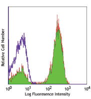
C57BL/6 mouse splenocytes were surface stained with mouse CD3. Cells were then intracellularly stained with Pax-5 (clone 1H9) Alexa Fluor® 647 (top) or rat IgG2a, κ Alexa Fluor® 647 isotype control (bottom). -

-

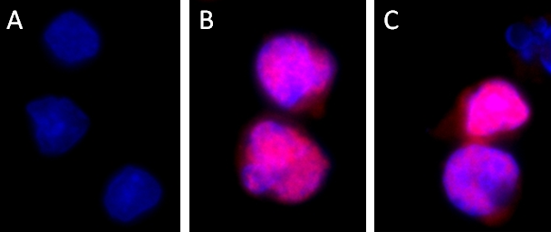
Mice were injected subcutaneously with sheep red blood cells in a volume of 25 µl per site on days 0 and 4 and harvested on day 11. Confocal image of C57BL/6 mouse lymph node acquired using the IBEX method of highly multiplexed antibody-based imaging: CD21 (purple) in Cycle 7 and Pax5 (cyan) in Cycle 8. Tissues were prepared using ~1% (vol/vol) formaldehyde and a detergent. Following fixation, samples are immersed in 30% (wt/vol) sucrose for cryoprotection. Images are courtesy of Drs. Andrea J. Radtke and Ronald N. Germain of the Center for Advanced Tissue Imaging (CAT-I) in the National Institute of Allergy and Infectious Diseases (NIAID, NIH).
| Cat # | Size | Price | Quantity Check Availability | Save | ||
|---|---|---|---|---|---|---|
| 649703 | 25 µg | £121 | ||||
| 649704 | 100 µg | £262 | ||||
Pax5, also known as BSAP (B cell specific activator protein), is a member of the paired box (Pax) family of transcription factors. PAX proteins are important regulators in early development, and alterations in the expression of their genes are thought to contribute to neoplastic transformation. Pax5 is the only member of the Pax family of transcription factors that is expressed in hematopoietic cells. During embryogenesis, Pax5 is transiently expressed in the brain of mice and in the mesencephalon and spinal cord of humans. Its expression is upregulated early in B cell development at the time of B cell commitment and is maintained throughout most subsequent stages. Suppression of Pax5 is essential for expression of Blimp-1 and the terminal differentiation of plasma cells. In the spleen, expression of Pax5 is higher in marginal zone B cells (B220+ CD21high CD23low) than in other B cells, especially the transition 1 stage (B220+ CD21- CD23-). In addition to its role in B cell development, Pax5 also affects VH-DJH heavy chain recombination as well as influencing the expression of many other B and non-B cell related proteins. Its expression has also been detected in developing CNS and testis and so the encoded protein may also play a role in neural development and spermatogenesis.
This gene is located at 9p13, which is involved in t(9;14)(p13;q32) translocations recurring in small lymphocytic lymphomas of the plasmacytoid subtype, and in derived large-cell lymphomas. This translocation brings the potent E-mu enhancer of the IgH gene into close proximity of the PAX5 promoter, suggesting that the deregulation of transcription of this gene contributes to the pathogenesis of these lymphomas. Alternatively spliced transcript variants encoding different isoforms have been described but their biological validity has not been determined.
Product Details
- Verified Reactivity
- Human, Mouse
- Antibody Type
- Monoclonal
- Host Species
- Rat
- Immunogen
- Recombinant mouse Pax-5 protein
- Formulation
- Phosphate-buffered solution, pH 7.2, containing 0.09% sodium azide.
- Preparation
- The antibody was purified by affinity chromatography and conjugated with Alexa Fluor® 647 under optimal conditions.
- Concentration
- 0.5 mg/ml
- Storage & Handling
- The antibody solution should be stored undiluted between 2°C and 8°C, and protected from prolonged exposure to light. Do not freeze.
- Application
-
ICFC - Quality tested
SB - Reported in the literature, not verified in house
- Recommended Usage
-
Each lot of this antibody is quality control tested by intracellular flow cytometry using our True-Nuclear™ Transcription Factor Staining Protocol. For flow cytometric staining, the suggested use of this reagent is ≤0.125 µg per million cells in 100 µl volume. It is recommended that the reagent be titrated for optimal performance for each application.
* Alexa Fluor® 647 has a maximum emission of 668 nm when it is excited at 633 nm / 635 nm.
Alexa Fluor® and Pacific Blue™ are trademarks of Life Technologies Corporation.
View full statement regarding label licenses - Excitation Laser
-
Red Laser (633 nm)
- Application Notes
-
The monoclonal 1H9 antibody recognizes both mouse and human Pax5.
Additional reported applications (for the relevant formats) include: Western blotting1,2, immunohistochemical staining of formalin-fixed sections, and spatial biology (IBEX)4,5.NOTE: For flow cytometric staining with this clone, True-Nuclear™ Transcription Factor Buffer Set (Cat. No. 424401) offers improved staining and is highly recommended over the Foxp3 Fix/Perm Buffer Set (Cat. No. 421403) and the Nuclear Factor Fixation and Permeabilization Buffer Set (Cat. No. 422601).
- Additional Product Notes
-
Iterative Bleaching Extended multi-pleXity (IBEX) is a fluorescent imaging technique capable of highly-multiplexed spatial analysis. The method relies on cyclical bleaching of panels of fluorescent antibodies in order to image and analyze many markers over multiple cycles of staining, imaging, and, bleaching. It is a community-developed open-access method developed by the Center for Advanced Tissue Imaging (CAT-I) in the National Institute of Allergy and Infectious Diseases (NIAID, NIH).
-
Application References
(PubMed link indicates BioLegend citation) - RRID
-
AB_2562424 (BioLegend Cat. No. 649703)
AB_2562425 (BioLegend Cat. No. 649704)
Antigen Details
- Structure
- 391 amino acids with predicted molecular weight of 42 kD
- Distribution
-
Nucleus
- Function
- Plays an important role in B-cell differentiation as well as neural development and spermatogenesis; involved in the regulation of the CD19 gene, a B-lymphoid-specific target gene
- Interaction
- Interacts with DAXX; binds DNA as a monomer, binds TLE4
- Cell Type
- B cells
- Biology Area
- Cell Biology, Immunology, Transcription Factors
- Molecular Family
- Nuclear Markers
- Antigen References
-
1. Liao F, et al. 1992. J. Immunol. 148:2909.
2. Nutt SL, et al. 1997. Genes Dev. 11:476.
3. Nera KP, et al. 2006. Immunity 24:283.
4. Fuxa M, et al. 2007. Immunity 178:3031. - Gene ID
- 5079 View all products for this Gene ID
- UniProt
- View information about Pax-5 on UniProt.org


Related FAQs
- If an antibody clone has been previously successfully used in IBEX in one fluorescent format, will other antibody formats work as well?
-
It’s likely that other fluorophore conjugates to the same antibody clone will also be compatible with IBEX using the same sample fixation procedure. Ultimately a directly conjugated antibody’s utility in fluorescent imaging and IBEX may be specific to the sample and microscope being used in the experiment. Some antibody clone conjugates may perform better than others due to performance differences in non-specific binding, fluorophore brightness, and other biochemical properties unique to that conjugate.
- Will antibodies my lab is already using for fluorescent or chromogenic IHC work in IBEX?
-
Fundamentally, IBEX as a technique that works much in the same way as single antibody panels or single marker IF/IHC. If you’re already successfully using an antibody clone on a sample of interest, it is likely that clone will have utility in IBEX. It is expected some optimization and testing of different antibody fluorophore conjugates will be required to find a suitable format; however, legacy microscopy techniques like chromogenic IHC on fixed or frozen tissue is an excellent place to start looking for useful antibodies.
- Are other fluorophores compatible with IBEX?
-
Over 18 fluorescent formats have been screened for use in IBEX, however, it is likely that other fluorophores are able to be rapidly bleached in IBEX. If a fluorophore format is already suitable for your imaging platform it can be tested for compatibility in IBEX.
- The same antibody works in one tissue type but not another. What is happening?
-
Differences in tissue properties may impact both the ability of an antibody to bind its target specifically and impact the ability of a specific fluorophore conjugate to overcome the background fluorescent signal in a given tissue. Secondary stains, as well as testing multiple fluorescent conjugates of the same clone, may help to troubleshoot challenging targets or tissues. Using a reference control tissue may also give confidence in the specificity of your staining.
- How can I be sure the staining I’m seeing in my tissue is real?
-
In general, best practices for validating an antibody in traditional chromogenic or fluorescent IHC are applicable to IBEX. Please reference the Nature Methods review on antibody based multiplexed imaging for resources on validating antibodies for IBEX.
Other Formats
View All Pax-5 Reagents Request Custom Conjugation| Description | Clone | Applications |
|---|---|---|
| Purified anti-Pax-5 | 1H9 | WB,ICC,IHC-P |
| Alexa Fluor® 647 anti-Pax-5 | 1H9 | ICFC,SB |
| Alexa Fluor® 488 anti-Pax-5 | 1H9 | ICFC |
| PE anti-Pax-5 | 1H9 | ICFC |
| PerCP/Cyanine5.5 anti-Pax-5 | 1H9 | ICFC |
| Alexa Fluor® 594 anti-Pax-5 | 1H9 | ICC |
| TotalSeq™-Bn1223 anti-Pax-5 | 1H9 | SB |
Customers Also Purchased
Compare Data Across All Formats
This data display is provided for general comparisons between formats.
Your actual data may vary due to variations in samples, target cells, instruments and their settings, staining conditions, and other factors.
If you need assistance with selecting the best format contact our expert technical support team.
-
Purified anti-Pax-5

Western blot analysis of Daudi (lane 1) and A20 (lane 2) cel... 
Ramos cells were fixed with 4% paraformaldehyde (PFA) for te... 
Tissue from human tonsil sections was fixed in 10% buffered ... -
Alexa Fluor® 647 anti-Pax-5

C57BL/6 mouse splenocytes were surface stained with mouse CD... 

Mice were injected subcutaneously with sheep red blood cells... -
Alexa Fluor® 488 anti-Pax-5

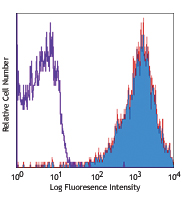
C57BL/6 mouse splenocytes were surface stained with mouse CD... 
-
PE anti-Pax-5

C57BL/6 mouse splenocytes were surface stained with mouse CD... 
-
PerCP/Cyanine5.5 anti-Pax-5

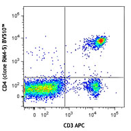
C57BL/6 mouse splenocytes were stained with mouse CD3 APC th... -
Alexa Fluor® 594 anti-Pax-5

Daudi cells were fixed with 4% paraformaldehyde (PFA) for 10... -
TotalSeq™-Bn1223 anti-Pax-5


Follow Us