 Login / Register


- Clone
- 3G8 (See other available formats)
- Regulatory Status
- RUO
- Workshop
- V NK80
- Other Names
- FcγRIII, Fc gamma receptor, Fc gamma receptor 3
- Isotype
- Mouse IgG1, κ
- Ave. Rating
- Submit a Review
- Product Citations
- publications

-

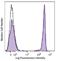
Human peripheral blood lymphocytes were stained with CD56 APC and CD16 (clone 3G8) Brilliant Violet 785™ (top) or mouse IgG1, κ Brilliant Violet 785™ isotype control (bottom). -

| Cat # | Size | Price | Quantity Check Availability | Save | ||
|---|---|---|---|---|---|---|
| 302045 | 25 tests | £155 | ||||
| 302046 | 100 tests | £275 | ||||
CD16 is known as low affinity IgG receptor III (FcγRIII). It is expressed as two distinct forms (CD16a and CD16b). CD16a (FcγRIIIA) is a 50-65 kD polypeptide-anchored transmembrane protein. It is expressed on the surface of NK cells, activated monocytes, macrophages, and placental trophoblasts in humans. CD16b (FcγRIIIB) is a 48 kD glycosylphosphatidylinositol (GPI)-anchored protein. Its extracellular domain is over 95% homologous to that of CD16a, and it is expressed specifically on neutrophils. CD16 binds aggregated IgG or IgG-antigen complex which functions in NK cell activation, phagocytosis, and antibody-dependent cell-mediated cytotoxicity (ADCC).
Product DetailsProduct Details
- Verified Reactivity
- Human, Cynomolgus, Rhesus
- Reported Reactivity
- African Green, Baboon, Capuchin Monkey, Chimpanzee, Common Marmoset, Pigtailed Macaque, Sooty Mangabey, Squirrel Monkey
- Antibody Type
- Monoclonal
- Host Species
- Mouse
- Immunogen
- Human PMN cells
- Formulation
- Phosphate-buffered solution, pH 7.2, containing 0.09% sodium azide and BSA (origin USA).
- Preparation
- The antibody was purified by affinity chromatography and conjugated with Brilliant Violet 785™ under optimal conditions.
- Concentration
- Lot-specific (to obtain lot-specific concentration and expiration, please enter the lot number in our Certificate of Analysis online tool.)
- Storage & Handling
- The antibody solution should be stored undiluted between 2°C and 8°C, and protected from prolonged exposure to light. Do not freeze.
- Application
-
FC - Quality tested
- Recommended Usage
-
Each lot of this antibody is quality control tested by immunofluorescent staining with flow cytometric analysis. For flow cytometric staining, the suggested use of this reagent is 5 µl per million cells in 100 µl staining volume or 5 µl per 100 µl of whole blood.
Brilliant Violet 785™ excites at 405 nm and emits at 785 nm. The bandpass filter 780/60 nm is recommended for detection, although filter optimization may be required depending on other fluorophores used. Be sure to verify that your cytometer configuration and software setup are appropriate for detecting this channel. Refer to your instrument manual or manufacturer for support. Brilliant Violet 785™ is a trademark of Sirigen Group Ltd.
Learn more about Brilliant Violet™.
This product is subject to proprietary rights of Sirigen Inc. and is made and sold under license from Sirigen Inc. The purchase of this product conveys to the buyer a non-transferable right to use the purchased product for research purposes only. This product may not be resold or incorporated in any manner into another product for resale. Any use for therapeutics or diagnostics is strictly prohibited. This product is covered by U.S. Patent(s), pending patent applications and foreign equivalents. - Excitation Laser
-
Violet Laser (405 nm)
- Application Notes
-
The 3G8 antibody clone blocks neutrophil phagocytosis and stimulates NK cell proliferation. It has been reported that this clone interacts with the FcγRIIa and FcγRIIIb receptors causing neutrophil activation and aggregation18. Due to this phenomenon staining in whole blood may cause a reduction in the number of granulocytes or alter their scatter profile.
Additional reported applications (for the relevant formats) include: immunohistochemical staining of acetone-fixed frozen tissue sections6, immunoprecipitation3, stimulation of NK cell proliferation4, blocking of phagocytosis5, and blocking of immunoglobulin binding to FcγRIII7,8. The Ultra-LEAF™ purified antibody (Endotoxin < 0.01 EU/µg, Azide-Free, 0.2 µm filtered) is recommended for functional assays (Cat. No. 302049, 302050, 302057, 302058). -
Application References
(PubMed link indicates BioLegend citation) -
- Knapp W, et al. Eds. 1989. Leucocyte Typing IV. Oxford University Press. New York.
- Schlossman S, et al. Eds. 1995. Leucocyte Typing V. Oxford University Press. New York.
- Edberg J, et al. 1997. J. Immunol. 159:3849. (IP)
- Hoshino S, et al. 1991. Blood 78:3232. (Stim)
- Tamm A, et al. 1996. Immunol. 157:1576. (Block)
- Da Silva DM, et al. 2001. Int. Immunol. 13:633. (IHC)
- Holl V, et al. 2004. J. Immunol. 173:6274. (Block)
- Hober D, et al. 2002. J. Gen. Virol. 83:2169. (Block)
- Brainard DM, et al. 2009. J. Virol. 83:7305. PubMed
- Smed-Sörensen A, et al. 2008. Blood 111:5037. (Block) PubMed
- Timmerman KL, et al. 2008. J. Leukoc. Biol. 84:1271. (FC) PubMed
- Yoshino N, et al. 2000. Exp. Anim. (Tokyo) 49:97. (FC)
- Rout N, et al. 2010. PLoS One 5:e9787. (FC)
- Kim WK, et al. 2006. Am. J. Pathol. 168:822. (FC)
- Boltz A, et al. 2011. J. Biol Chem. 286:21896. PubMed
- Wu Z, et al. 2013. J. Virol. 87:7717. PubMed
- Peterson VM, et al. 2017. Nat. Biotechnol. 35:936. (PG)
- Vossebeld PJ, et al. 1997. Biochem J. 323:87-94 (Stim)
- Product Citations
-
- RRID
-
AB_2561367 (BioLegend Cat. No. 302045)
AB_2563803 (BioLegend Cat. No. 302046)
Antigen Details
- Structure
- Ig superfamily, transmembrane form (50-65 kD) or GPI-linked form (48 kD)
- Distribution
-
NK cells, activated monocytes, macrophages, neutrophils
- Function
- Low affinity IgG Fc receptor, phagocytosis, ADCC
- Ligand/Receptor
- Aggregated IgG, IgG-antigen complex
- Cell Type
- Dendritic cells, Macrophages, Monocytes, Neutrophils, NK cells
- Biology Area
- Immunology, Innate Immunity
- Molecular Family
- CD Molecules, Fc Receptors
- Antigen References
-
1. Fleit H, et al. 1982. P. Natl. Acad. Sci. USA 79:3275.
2. Stroncek D, et al. 1991. Blood 77:1572.
3. Wirthmueller U, et al. 1992. J. Exp. Med. 175:1381. - Gene ID
- 2214 View all products for this Gene ID
- UniProt
- View information about CD16 on UniProt.org
Related Pages & Pathways
Pathways


Related FAQs
- Is our human Trustain FcX™ (cat# 422302) compatible with anti human CD16, CD32 and CD64 clones 3G8, FUN-2 and 10.1 respectively?
-
Yes
Other Formats
View All CD16 Reagents Request Custom ConjugationCustomers Also Purchased
Compare Data Across All Formats
This data display is provided for general comparisons between formats.
Your actual data may vary due to variations in samples, target cells, instruments and their settings, staining conditions, and other factors.
If you need assistance with selecting the best format contact our expert technical support team.
-
APC anti-human CD16

Human peripheral blood lymphocytes stained with 3G8 APC -
Biotin anti-human CD16

Human peripheral blood lymphocytes stained with biotinylated... -
FITC anti-human CD16

Human peripheral blood lymphocytes stained with 3G8 FITC -
Brilliant Violet 711™ anti-human CD16

Human peripheral blood lymphocytes were stained with CD16 (c... -
PE anti-human CD16

Human peripheral blood lymphocytes stained with 3G8 PE 
Human peripheral blood was stained with CD16 (clone 3G8) PE ... -
PE/Cyanine5 anti-human CD16

Human peripheral blood lymphocytes stained with 3G8 PE/Cyani... -
Purified anti-human CD16

Human peripheral blood lymphocytes stained with purified 3G8... -
APC/Cyanine7 anti-human CD16

Human peripheral blood lymphocytes stained with CD56 (NCAM) ... -
PE/Cyanine7 anti-human CD16
Human peripheral blood lymphocytes stained with 3G8 PE/Cyani... -
Alexa Fluor® 488 anti-human CD16

Human peripheral blood lymphocytes stained with 3G8 Alexa Fl... -
Alexa Fluor® 647 anti-human CD16

Human peripheral blood lymphocytes stained with 3G8 Alexa Fl... -
Pacific Blue™ anti-human CD16

Human peripheral blood lymphocytes stained with 3G8 Pacific ... -
Alexa Fluor® 700 anti-human CD16

Human peripheral blood lymphocytes stained with 3G8 Alexa fl... -
PerCP/Cyanine5.5 anti-human CD16

Human peripheral blood lymphocytes were stained with CD56 AP... -
PerCP anti-human CD16

Human peripheral blood lymphocytes stained with 3G8 PerCP -
Brilliant Violet 421™ anti-human CD16

Human peripheral blood lymphocytes were stained with CD16 (c... -
Brilliant Violet 570™ anti-human CD16

Human peripheral blood lymphocytes were stained with CD16 (c... -
Brilliant Violet 605™ anti-human CD16

Human peripheral blood lymphocytes were stained with CD16 (c... -
Brilliant Violet 650™ anti-human CD16

Human peripheral blood lymphocytes were stained with CD16 (c... -
Brilliant Violet 785™ anti-human CD16

Human peripheral blood lymphocytes were stained with CD56 AP... 
-
Brilliant Violet 510™ anti-human CD16

Human peripheral blood lymphocytes were stained with CD16 (c... -
Ultra-LEAF™ Purified anti-human CD16

Human peripheral blood lymphocytes stained with Ultra-LEAF™ ... -
Purified anti-human CD16 (Maxpar® Ready)

Human PBMCs stained with 152Sm-anti-CD13 (WM15) and 148Nd-an... -
PE/Dazzle™ 594 anti-human CD16

Human peripheral blood lymphocytes were stained with CD16 (c... -
APC/Fire™ 750 anti-human CD16

Human peripheral blood lymphocytes were stained with CD56 FI... 
-
PE anti-human CD16

Typical results from human peripheral blood lymphocytes stai... -
APC anti-human CD16

Typical results from human peripheral blood lymphocytes stai... -
Pacific Blue™ anti-human CD16

Typical results from human peripheral blood lymphocytes sta... -
PE/Dazzle™ 594 anti-human CD16

Typical results from human peripheral blood lymphocytes stai... -
TotalSeq™-A0083 anti-human CD16
-
TotalSeq™-B0083 anti-human CD16
-
TotalSeq™-C0083 anti-human CD16
-
PE/Cyanine7 anti-human CD16
Typical results from human peripheral blood lymphocytes stai... -
PE/Fire™ 640 anti-human CD16

Human peripheral blood lymphocytes were stained with CD56 (N... -
FITC anti-human CD16

Typical results from human peripheral blood lymphocytes stai... -
Spark YG™ 581 anti-human CD16

Human peripheral blood lymphocytes were stained with anti-hu... -
APC/Fire™ 750 anti-human CD16

Typical results from human peripheral blood lymphocytes stai... -
TotalSeq™-D0083 anti-human CD16
-
APC/Fire™ 810 anti-human CD16

Human peripheral blood lymphocytes were stained with anti-hu... -
GMP APC anti-human CD16

Typical results from human peripheral blood lymphocytes stai... -
GMP PE/Dazzle™ 594 anti-human CD16

Typical results from human peripheral blood lymphocytes stai... -
GMP PE anti-human CD16

Typical results from human peripheral blood lymphocytes stai... -
Spark Red™ 718 anti-human CD16

Human peripheral blood lymphocytes were stained with anti-hu... -
GMP Pacific Blue™ anti-human CD16

Typical results from human peripheral blood lymphocytes stai... -
GMP FITC anti-human CD16

Typical results from human peripheral blood lymphocytes stai... -
Spark Blue™ 515 anti-human CD16

Human peripheral blood lymphocytes were stained with anti-hu... -
Spark UV™ 387 anti-human CD16

Human peripheral blood lymphocytes were stained with anti-hu... -
PerCP/Cyanine5.5 anti-human CD16

Typical results from human peripheral blood lymphocytes stai... -
GMP PE/Cyanine7 anti-human CD16

Typical results from human peripheral blood lymphocytes stai... -
GMP APC/Fire™ 750 anti-human CD16

Typical results from human peripheral blood lymphocytes stai... -
Brilliant Violet 750™ anti-human CD16

Human peripheral blood lymphocytes were stained with anti-hu... -
Spark Blue™ 550 anti-human CD16

Human peripheral blood lymphocytes were stained with anti-hu... -
GMP PerCP/Cyanine5.5 anti-human CD16

Typical results from human peripheral blood lymphocytes stai... -
Spark Violet™ 500 anti-human CD16

Human peripheral blood lymphocytes were stained with anti-hu... -
Spark Blue™ 574 anti-human CD16 (Flexi-Fluor™)
-
Spark PLUS UV™ 395 anti-human CD16

Human peripheral blood lymphocytes were stained with anti-hu...


Follow Us