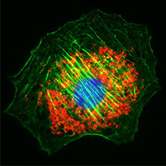
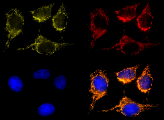
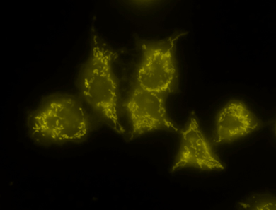
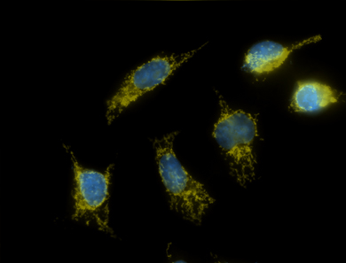
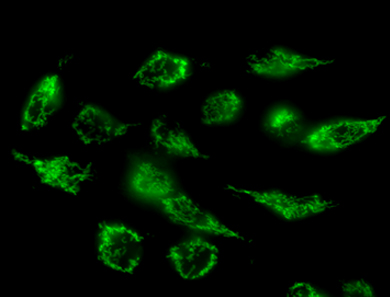
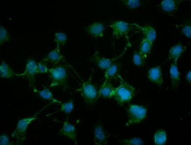
MitoSpy™ Orange, MitoSpy™ Red, and MitoSpy™ NIR can also be used as an indicator of cellular health. When the mitochondria are actively respiring, there is a potential difference across the mitochondrial membrane that is called membrane polarization. If a cell is unhappy due to apoptosis or cell death, this probe will not be strongly attracted to the mitochondria of that cell. In the image below, cells positive for MitoSpy™ Orange, MitoSpy™ Red, MitoSpy™ NIR are alive and healthy, while Annexin V positive events are at the beginning stages of apoptosis. In this flow cytometry application, the MitoSpy™ Orange, MitoSpy™ Red, and MitoSpy™ NIR should not be fixed prior to analysis since there is a substantial loss of reagent with fixation. An apoptotic phenotype is indicated by a low or negative MitoSpy™ Orange, MitoSpy™ Red, or MitoSpy™ NIR signal. If reagent is lost with fixation, the resulting loss of signal intensity will confound the ability to accurately phenotype apoptosis. Fixation of MitoSpy™ Orange and MitoSpy™ Red is only applicable for subcellular localization in imaging applications. Fixation is not recommended at all with MitoSpy™ NIR.
 Login/Register


Follow Us