Growth factors are signaling molecules that regulate a number of critical cellular processes including proliferation, differentiation, migration, and development. Growth factors include both cytokines and hormones, which often initiate downstream signaling through binding to receptor tyrosine kinases on cellular surfaces. BioLegend offers a wide range of antibodies and recombinant proteins to study growth factors in a variety of applications including bioassays, ELISAs, multiplexing, Western blotting, flow cytometry and microscopy. View the sections below to learn more about the different growth factor families.


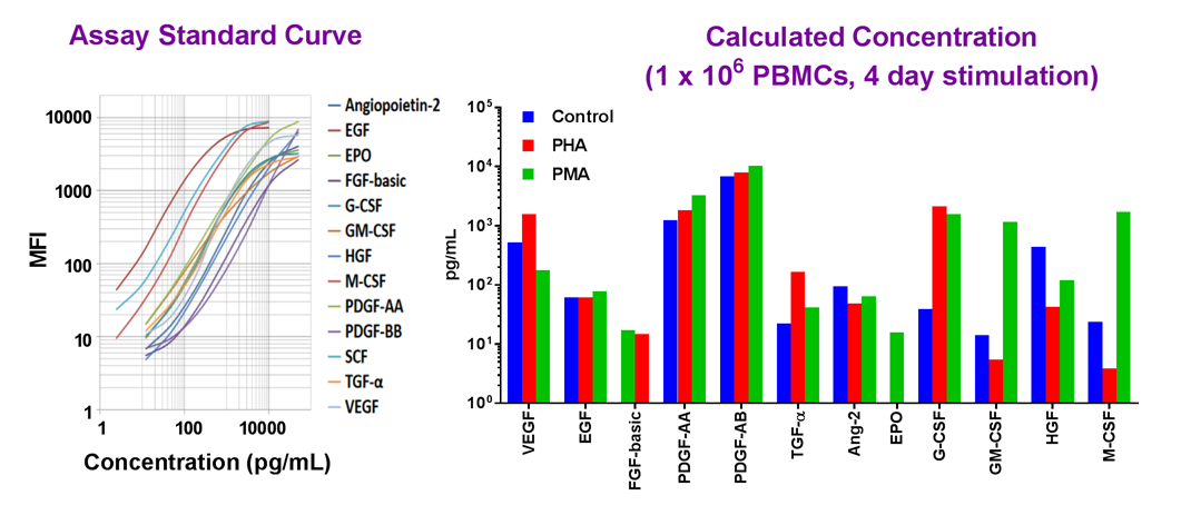
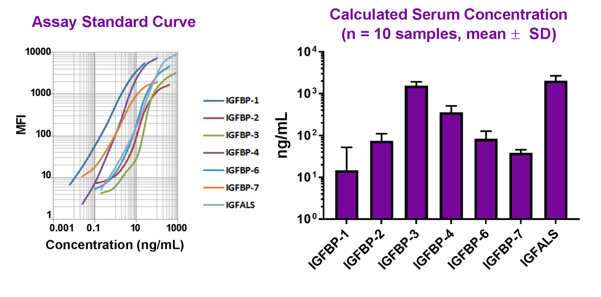
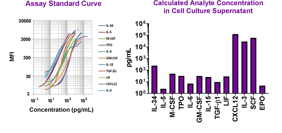
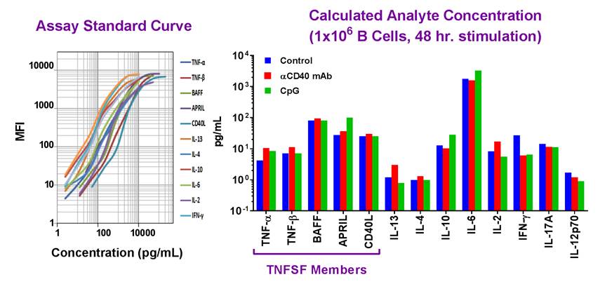
Simultaneously measure 13 human growth factors with our LEGENDplex™ bead-based immunoassay panel.

BMP Family

Bone morphogenetic proteins (BMPs) belong to the TGF-β superfamily and play a role in embryonic development, particularly during heart, neural, and cartilage development. As their name suggests, they also regulate bone organogenesis. Currently, around 20 bone morphophogenetic proteins (BMPs) have been identified and are typically characterized into 4 distinct subfamilies based on structural similarities. BMPs bind to serine/threonine kinase receptors which are usually separated into both type I and type II subtypes. Following binding to BMPs, BMP receptors activate SMAD proteins which translocate to the nucleus and regulate transcription of target genes. Cross-talk between BMP signaling and other pathways such as MAPK, Notch, Wnt, and Hedgehog pathways contribute to osteogenesis and other developmental pathways.

Featured Data

Ultra-LEAF™ Purified anti-human BMP9

Recombinant human BMP9 (Cat. No. 553102) induces alkaline phosphatase in ATDC5 mouse chondrogenic cell line in a dose dependent manner (black circles). Anti-human BMP9 monoclonal antibody (clone A16035H, purple circles) neutralizes the production of alkaline phosphatase in ATDC5 cells induced by BMP9 (2 ng/mL). The ND50= 0.05 - 0.25 μg/mL.

EGF Family

Epidermal growth factor (EGF) family members play a critical role in cell proliferation, differentiation, and survival. Other than EGF, the first identified family member, the family contains HB-EGF, TGF-α, amphiregulin, epiregulin, epigen, betacellulin and neuregulins. All of the family members contain 6 cysteine residues which form 3 disulfide bonds generating a structural loop required for binding EGF receptors (EGFRs). EGFRs are receptor tyrosine kinases that, when activated, dimerize and initiate downstream MAPK, AKT, and JNK signaling pathways. EGF and EGFRs can be found upregulated in a number of different cancer types.

Featured Data

Purified anti-EGF Antibody


Immunoprecipitation/Western blot analysis of 200ng recombinant EGF. Lane 1 was immunoprecipitated with mouse IgG control antibody, lane 2 was immunoprecipitated with EGF antibody (clone M1401E03), and lane 3 was loaded with 25ng recombinant human EGF. Western blot analysis was performed using goat anti-human EGF antibody followed by HRP-anti-goat IgG.

LEGENDplex™ Human Growth Factor Panel

Human PBMCs (1x10⁶ cells/mL) were cultured under various conditions (PHA, 10 μg/mL; PMA, 50 ng/mL; Control, no treatment). Supernatants were collected after 4 days then assayed with the LEGENDplex™ Human Growth Factor Panel (Cat. No. 740180).

FGF Family

Fibroblast growth factor (FGF) family members play key roles in signaling during development, regulating diverse pathways such as proliferation, survival, migration, differentiation and organogenesis. In adults, FGF signaling is induced in injury and plays a role in wound healing. Expressed in almost all tissues, the FGF family includes 23 members which bind to four distinct tyrosine kinase receptors designated FGFR1-4 and function in downstream signaling pathways including RAS-MAPK, PI3K-AKT, PLCγ, and STAT. In addition, there are non-signaling FGF molecules which regulate intracellular voltage gated sodium channels.

Featured Data

Purified anti-FGF-basic Antibody


U87-MG cells were fixed with 4% paraformaldehyde (PFA) for ten minutes, permeabilized with 0.5% Triton X-100 for ten minutes, and blocked with 5% FBS for 30 minutes. Then the cells were intracellularly stained with 2 µg/mL purified anti-FGF-basic (clone A15021A) antibody for two hours at room temperature followed by Alexa Fluor® 594 conjugated goat anti-mouse IgG (red) for one hour at room temperature. Actin filaments were labeled with Alexa Fluor® 488 Phalloidin (green). Nuclei were counterstained with DAPI (blue). The image was captured with a 60X objective.

LEGENDplex™ Human Growth Factor Panel

Human PBMCs (1x10⁶ cells/mL) were cultured under various conditions (PHA, 10 μg/mL; PMA, 50 ng/mL; Control, no treatment). Supernatants were collected after 4 days then assayed with the LEGENDplex™ Human Growth Factor Panel (Cat. No. 740180).

Hedgehog Family

The Hedgehog protein family regulates early development and stem cell maintenance and differentiation. In adult tissues, they are important in tissue remodeling and aberrant regulation of this pathway is associated with carcinogenesis. In vertebrates, the Hedgehog family contains sonic hedgehog (SHH), Indian hedgehog (IHH) and Desert hedgehog (DHH). Hedgehog proteins are cleaved to yield an N-terminal fragment which is important for signaling and a C-terminal fragment which is glycosylated and is active in precursor processing. The C-terminal domain mediates the covalent attachment of a cholesterol moiety to the N-terminal restricting it to the cell surface. In conjunction with the Hedgehog interaction protein (HIP), hedgehog proteins bind to the Patched receptor, which associates with the Smoothened receptor, to activate transcription of target genes.

Featured Data

Recombinant Human Sonic Hedgehog (carrier-free)


Recombinant Human SHH induces C3H10T1/2 differentiation in the presence of hBMP9 (20 ng/mL) as measured by alkaline phosphatase production. Alkaline phosphatase activity was measured using p-nitrophenyl phosphate.

Recombinant Mouse IHH (carrier-free)


Recombinant mouse IHH (Cys28/(Ile-Ile)-Gly202) induces C3H10T1/2 cell differentiation in the presence of hBMP9 (20 ng/mL) as measured by alkaline phosphatase production. Alkaline phosphatase activity was measured using p-nitrophenyl phosphate.

IGF Family

The insulin-growth factors (IGF) family contains three members (IGF-I, IGF-II, and IGF-III) and shares a structural similarity with insulin. IGFs act as a hormone and are important during growth and development and have anabolic effects in adult tissues. The IGF family binds to one of two IGF tyrosine kinase receptors, and this signaling cascade regulates proliferation, survival, differentiation, and migration. There are seven IGF binding proteins (IGFBP) which serve to both regulate the availability of IGF family members and extend the half-life of the IGF peptides. Additionally, as the binding proteins bind to the IGFs with different affinities they also regulate interactions with the IGF receptors and can modulate their actions.

Featured Data

Ultra-LEAF™ Purified anti-human IGFBP-4 Antibody


Recombinant human IGFBP-4 binds human IGF-I in a dose-dependent manner (black circles). The binding of human IGF-I to human IGFBP-4 is blocked (purple circles) by increasing concentrations of anti-human IGFBP-4 monoclonal antibody (Clone A15038E). The ND50 is typically 4 - 12 μg/mL.


LEGENDplex™ Human IGFBP Panel

Normal human serum samples from 10 different donors were tested for endogenous levels of human IGFBP proteins using the LEGENDplex™ Human IGFBP Panel (Cat. No. 740468).

Neurotrophic Family

Neurotrophin growth factors regulate the development, growth, and survival of neurons specifically in regulating injury induced neuron regeneration, differentiation of neuronal populations in the peripheral and central nervous system, axonal growth, and modulating dendritic growth and morphology. These growth factors share a tertiary-folded structure containing a cysteine-knot domain. Though neurotrophins function in a specific cell type, the signaling mechanism is similar to other growth factor families as they bind tyrosine kinase receptors which autophosphorylate initiating a phosphorylation cascade. Dysregulation of neurotrophins is related to neurological disorders such as Alzheimer's disease.

Featured Data

Ultra-LEAF™ Purified anti-mouse Thrombopoietin Antibody


Recombinant mouse TPO induces proliferation of human megakaryocytic leukemic MO7e cells in a dose dependent manner (black circles). Anti-mouse TPO monoclonal antibody (clone A15139F, purple circles) neutralizes the proliferation of MO7e cells induced by recombinant mouse TPO (1 ng/ml). The ND50 = 0.04 – 0.2 μg/ml.


LEGEND MAX™ Mouse LIF ELISA kit


Lungs were harvested from a mouse and dissected into small pieces (~1 mm3) and cultured in 10% FBS/RPMI. Supernatants were removed at 1, 2, 3, and 7 days and LIF concentrations were quantified using the LEGEND MAX™ Mouse LIF ELISA Kit (Cat. No. 445107).

PDGF Family

The platelet-derived growth factor (PDGF) family is defined by 5 different isotypes consisting of dilsulfide-linked homo- or hetero-dimers of four poly peptides known as the A, B, C, and D chains. Like the VEGF family, PDGFs contain a cysteine-knot domain. PDGF-A and –B chains must undergo intracellular activation while PDGF-C and –D are secreted before being processed by extracellular proteases. PDGFs are mitogenic, function to direct cell migration, and are typically found in neural or mesenchymal cells. Additionally, PDGFs are synthesized, stored, and released by platelets upon activation. PDGF receptors are dimeric receptors consisting of two tyrosine-kinase receptor subunits and upon surface binding signal through a phosphorylation cascade.

Featured Data

LEGENDplex™ Human Growth Factor Panel


Human PBMCs (1x10⁶ cells/mL) were cultured under various conditions (PHA, 10 μg/mL; PMA, 50 ng/mL; Control, no treatment). Supernatants were collected after 4 days then assayed with the LEGENDplex™ Human Growth Factor Panel (Cat. No. 740180).

Stem Cell Proteins

Certain growth factors, sometimes referred to as hematopoietic growth factors, regulate the proliferation and survival of stem cells as well as influence differentiation of hematopoietic stem cells to various lineages. These growth factors are often found in the bone marrow niche and can be produced by fibroblasts, endothelial cells, and mature lymphocytes, monocytes and macrophages. As these cytokines directly influence differentiation into particular cell types, recombinant growth factors have been used in clinical settings. For example, exogenously administered EPO can stimulate erythropoiesis and is used to help treat anemias. Other stem cell growth factors such as G-CSF and GM-CSF are often used during bone marrow transplantation to stimulate the donor pool of peripheral blood progenitors.

Featured Data

Alexa Fluor® 594 anti-LIF Antibody


HeLa cells were fixed with ice cold methanol for five minutes and blocked with 5% FBS for 30 minutes. Then, the cells were intracellularly stained with 2 μg/ml Alexa Fluor® 594 conjugated (red) LIF Antibody (clone M1506B09) in a blocking buffer overnight at 4°C. Nuclei were counterstained with DAPI (blue). The image was captured with a 60X objective.

LEGENDplex™ Mouse HSC Panel


Head tissue from E10.5 mouse embryos (n = 6) was digested and then sorted using FACS to isolate endothelial cells (CD31posCD41negCD45neg). Cells were then cultured with OP9-DL1 cells, a bone-marrow derived stromal cell line that ectopically expresses the Notch ligand, Delta-like 1 (DL1). This in vitro culture system allows for the commitment, differentiation, and proliferation of T cells from hemogenic endothelial cells. Cells were co-cultured for 7 days, and then supernatants were assayed using the LEGENDplex™ Mouse HSC panel (Cat. No. 740676). Data courtesy of Dr. Zhuan Li, MRC Centre for Inflammation Research at the University of Edinburgh.

TGF Family

The transforming growth factor (TGF) superfamily contains over 30 proteins, including TGFβs, BMPs and activins. The TGF family members, particularly the earliest member TGF-β, have been well-studied for their role in immunoregulation including roles in inflammation and response to infection. Additionally, TGFs play an important role in development and tissue homeostasis. TGF family members typically exist as either homo- or heterodimers and are proteolytically cleaved prior to activation and subsequent receptor binding. TGFs bind to what is designated as type I and type II receptor complexes. TGF receptors typically signal through SMAD proteins to regulate transcription of target genes in the nucleus; however, TGF family members can also activate p38, JNK, ERK-MAPK, and PI3K-AKT signaling pathways. As TGFs regulate the immune system and proliferation/growth pathways, they can both suppress and support tumor growth, depending on the cell context.

Featured Data

Ultra-LEAF™ Purified anti-Activin A antibody


Recombinant human Activin A (grey triangles) inhibits the proliferation of mouse plasmacytoma cell line MPC 11. Anti-human Activin A antibody (clone A15095A, purple squares) inhibits the effect of recombinant Activin A (at 30 ng/ml) in MPC 11 cells in a dose dependent manner. The ND50 = 0.35 - 1.05 μg/ml.

TNF Family

The Tumor necrosis factor (TNF) superfamily is one of the largest growth factor families as it consists of approximately 19 ligands which bind to approximately 29 receptors. The TNF family plays important roles in regulating the immune response and hematopoiesis. While many TNF family members are membrane-bound ligands which act locally, they can also be shed from the membrane and the soluble ligands can act as either agonists or antagonists. Once bound to a TNF receptor, TNF growth factors mediate apoptosis, particularly those ligands which bind receptors containing a death domain, or regulate proliferation by activating pathways such as the NF-κB, JNK, or MAPK signaling pathways. Like most pathways involved in apoptosis and proliferation, alterations with the TNF superfamily are associated with a number of disease states including tumorigenesis and autoimmune diseases.

Featured Data

LEGENDplex™ Human B Cell Panel


Human B Cells (1 x 10⁶ cells/mL) were isolated and cultured under various conditions (activating CD40 mAb, 5 μg/mL; CpG, 5 μg/mL; Control, no treatment). Supernatants were collected after 48 hours and assayed with the LEGENDplex™ Human B Cell Panel (Cat. No. 740526). TNFSF Members are denoted by purple bracket.

PE anti-human CD357 (GITR) Antibody


Human peripheral blood lymphocytes were activated for 3 days with PHA, and then stained with CD3 Alexa Fluor® 647 and CD357 (GITR) (clone 108-17) PE (top) or mouse IgG2a, κ PE isotype control (bottom).

VEGF Family

Vascular endothelial growth factors (VEGFs) are widely expressed mitogens, particularly important in vascular endothelial cells where they play a critical role in regulating angiogenesis and permeability of capillary blood vessels. Cytokines, hypoxia, and oncogenes can all induce VEGF expression. Like PDGF family members, they contain eight cysteine residues which form what is known as a cysteine-knot structure. There are five main members of the VEGF family: VEGF-A, VEGF-B, VEGF-C, VEGF-D, and placental growth factor (PGF). Additionally, VEGF-A (commonly referred to as VEGF) can be alternatively spliced to produce a number of different isoforms. In general, VEGF family members bind to receptor tyrosine kinases (VEGFRs) on the cell surface where the receptors dimerize and become activated through transphosphorylation. VEGF can also bind to neuropilins on neurons where they stimulate angiogenesis.

Featured Data

Ultra-LEAF™ Purified anti-VEGF-121 antibody


Recombinant human VEGF-121 stimulates proliferation of HUVEC in a dose-dependent manner (dark purple squares). Proliferation induced by recombinant human VEGF-121 (15 ng/mL) is neutralized (purple circles) by increasing concentrations of anti-VEGF-121 (clone A15136B) antibody. The ND50 is typically 25 - 100 ng/mL.

LEGENDplex™ Human Growth Factor Panel


Human PBMCs (1x10⁶ cells/mL) were cultured under various conditions (PHA, 10 μg/mL; PMA, 50 ng/mL; Control, no treatment). Supernatants were collected after 4 days then assayed with the LEGENDplex™ Human Growth Factor Panel (Cat. No. 740180).

Other growth factors

Other growth factors do not fall under a specific family name but like all growth factors, they typically exist in a soluble form and bind to transmembrane receptors initiating downstream signaling cascades. Other than influencing proliferation, differentiation and immune responses, these growth factors have specific functions in certain cell types. For example, prolactin is best known for its role in enabling lactation and asprosin regulates metabolic pathways and promotes glucose release in hepatocytes.

Featured Data

Ultra-LEAF™ Purified anti-human S100A8/A9 Heterodimer Antibody


Recombinant human S100A8/A9 heterodimer stimulates proliferation of astrocytes in a dose-dependent manner (black circles). Proliferation induced by human S100A8/A9 heterodimer is neutralized (purple circles) by increasing concentrations of anti-human S100A8/A9 monoclonal antibody (Clone A15015B). The ND50 is typically 2 - 8 μg/mL.

ProductsHere

Login/Register
Remember me
Forgot your password? Reset Password
Request an Account