 Login / Register


- Regulatory Status
- RUO
- Other Names
- SAv-Brilliant Violet 421™
- Ave. Rating
- Submit a Review
- Product Citations
- publications

-

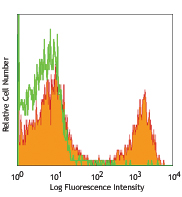
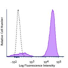
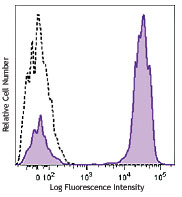
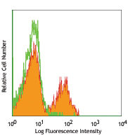
Human peripheral blood lymphocytes were stained with biotinylated CD3 (filled histogram) or mouse IgG1 isotype control (open histogram), followed with SAV-Brilliant Violet 421™.
| Cat # | Size | Price | Quantity Check Availability | Save | ||
|---|---|---|---|---|---|---|
| 405226 | 100 µL | £133 | ||||
| 405225 | 100 µg | £317 | ||||
Streptavidin binds to biotin with high affinity. Streptavidin-Brilliant Violet 421™ is useful for detecting biotinylated antibodies. The excitation of Brilliant Violet 421™ by 405 nm laser light induces a fluorescence maximum emission of 421 nm.
Product DetailsProduct Details
- Verified Reactivity
- Human, Mouse, Rat, All Species
- Formulation
-
Cat. No. 405225: Phosphate-buffered solution, pH 7.2, containing 0.09% sodium azide.
Cat. No. 405226: Phosphate-buffered solution, pH 7.2, containing 0.09% sodium azide and BSA (origin USA). - Preparation
- Streptavidin was conjugated with Brilliant Violet 421™ under optimal conditions.
- Concentration
-
405225 is bottled at 0.5 mg/ml.
405226 is bottled at 0.1 mg/ml.
(concentration relates to the Streptavidin only component of the conjugate) - Storage & Handling
- The solution should be stored undiluted between 2°C and 8°C, and protected from prolonged exposure to light. Do not freeze.
- Application
-
FC, ICFC - Quality tested
ICC, IHC-F, IHC-P - Recommended Usage
-
Each lot of this Streptavidin-Brilliant Violet 421™ is quality control tested by immunofluorescent staining with flow cytometric analysis. The concentration provided is based upon molecular mass of streptavidin independent of any additional molecular mass that might be added by the Brilliant Violet 421™ conjugation. For immunofluorescent staining, we recommend using Cat. No. 405226 at dilutions ≤ 0.015 µg in 100 µL staining volume per million cells. For applications requiring high concentration of streptavidin reagent such as tetramer labeling, we recommend using Cat. No. 405225. It is recommended that the reagent be titrated for optimal performance for each application.
* Brilliant Violet 421™ excites at 405 nm and emits at 421 nm. The standard bandpass filter 450/50 nm is recommended for detection. Brilliant Violet 421™ is a trademark of Sirigen Group Ltd.
Learn more about Brilliant Violet™.
This product is subject to proprietary rights of Sirigen Inc. and is made and sold under license from Sirigen Inc. The purchase of this product conveys to the buyer a non-transferable right to use the purchased product for research purposes only. This product may not be resold or incorporated in any manner into another product for resale. Any use for therapeutics or diagnostics is strictly prohibited. This product is covered by U.S. Patent(s), pending patent applications and foreign equivalents. - Excitation Laser
-
Violet Laser (405 nm)
- Application Notes
-
Streptavidin-Brilliant Violet 421™ is useful as a second step reagent for indirect immunofluorescent staining when used in conjunction with biotinylated primary antibodies. The average molecular weight of Streptavidin-Brilliant Violet 421™ is 340 kD and Streptavidin alone is 52 kD.
- Additional Product Notes
-
View more applications data using this product in immunofluorescence microscopy and labeling tetramers in flow cytometry in our Scientific Poster Library.
-
Application References
(PubMed link indicates BioLegend citation) -
- Kim G, et al. 2013. J. Immunol. 190:1510. PubMed.
- Giltiay NV, Et al. 2013. J. Immunol. 191:2155. PubMed
- Sugiyama D, et al. 2013. PNAS. 110:17945. PubMed
- Scally SW, et al. 2013. J. Exp Med. 210:2569. PubMed
- Lyngaa R, et al. 2014. Clin Cancer Res. 20:1768. PubMed
- Higdon LE, et al. 2014. PNAS. 111:5652. PubMed
- Friedman RS, et al. 2014. PNAS. 111:9223. PubMed
- van de Weijer ML, et al. 2014. Nat Commun. 5:3832. PubMed
- Mise-Omata S, et al. 2014. Int Immunol. PubMed
- Choo JA, et al. 2014. J Virol. 88:10613. PubMed
- Xing YL, et al. 2014. J Neurosci. 34:14128. PubMed
- Luetke-Eversloh M, et al. 2014. PLoS Pathog. 10:1004441. PubMed
- Poh Cm, et al. 2014. Infect Immun. 82:4854. PubMed
- Cabrera-Perez J, et al. 2015. J Immunol. 194:1609. PubMed
- Lucas A, et al. 2015. PLoS One. 10:117160. PubMed
- Krishnaswamy JK, et al. 2015. PNAS. 112:3056. PubMed
- Rampuria P, et al. 2015. Int Immunol. 27:253. PubMed
- Product Citations
-
Antigen Details
- Gene ID
- NA
- UniProt
- View information about Biotin on UniProt.org


Related FAQs
- What is the F/P ratio range of our BV421™ format antibody reagents?
-
It is lot-specific. On average it ranges between 2-4.


Follow Us