 Login / Register


- Clone
- W6/32 (See other available formats)
- Regulatory Status
- RUO
- Other Names
- Major Histocompatibility Class I, MHC class I
- Isotype
- Mouse IgG2a, κ
- Ave. Rating
- Submit a Review
- Product Citations
- publications

-

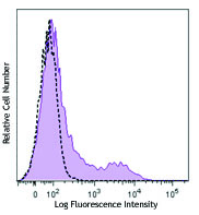
Human peripheral blood lymphocytes were stained with purified anti-HLA-A,B,C (clone W6/32, filled histogram) or purified mouse IgG2a, κ isotype control (open histogram), followed by goat-anti-mouse IgG FITC. -

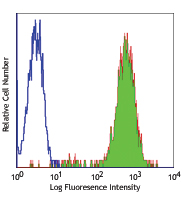
HEK293 cells were transfected with RelA or empty vector and 24hrs later cell extracts harvested using a 1% CHAPS lysis buffer. Extracts were resolved by non-denaturing, non-reducing electrophoresis, transferred to nitrocellulose, and probed with a 1:500 dilution purified W6/32 . Proteins were visualized using a goat anti-mouse secondary antibody conjugated to HRP and a chemiluminescence detection system. These data document that MHC class I was upregulated in cells constitutively expressing RelA. (Data was provided by Dr. Ezra Burstein, University of Michigan Medical School, Ann Arbor, MI).
| Cat # | Size | Price | Quantity Check Availability | Save | ||
|---|---|---|---|---|---|---|
| 311402 | 100 µg | £61 | ||||
MHC class I antigens associated with β2-microglobulin are expressed by all human nucleated cells. MHC class I molecules are involved in presentation of antigens to CD8+ T cells. They play an important role in cell-mediated immune responses and tumor surveillance.
Product DetailsProduct Details
- Verified Reactivity
- Human, Cynomolgus, Rhesus
- Reported Reactivity
- African Green, Baboon, Cat, Cow, Chimpanzee
- Antibody Type
- Monoclonal
- Host Species
- Mouse
- Formulation
- Phosphate-buffered solution, pH 7.2, containing 0.09% sodium azide.
- Preparation
- The antibody was purified by affinity chromatography.
- Concentration
- 0.5 mg/ml
- Storage & Handling
- The antibody solution should be stored undiluted between 2°C and 8°C.
- Application
-
FC - Quality tested
Activ, Block, IHC-F, IP, WB - Reported in the literature, not verified in house - Recommended Usage
-
Each lot of this antibody is quality control tested by immunofluorescent staining with flow cytometric analysis. For flow cytometric staining, the suggested use of this reagent is ≤ 2.0 µg per 106 cells in 100 µl volume or 100 µl of whole blood. It is recommended that the reagent be titrated for optimal performance for each application.
- Application Notes
-
Clone W6/32 recognizes residues in the N terminus of the human ß2-microglobulin molecule21.
Additional reported applications (for the relevant formats) include: immunoprecipitaton2, Western blotting (non-reducing)3, immunohistochemical staining of acetone-fixed frozen tissue sections4,5, blocking6,7, inhibition of NK cell-mediated lysis10, and activation8,9. Clone W6/32 has been reported not to be suitable for immunohistochemistry on paraffin sections17. The LEAF™ purified antibody (Endotoxin < 0.1 EU/µg, Azide-Free, 0.2 µm filtered) is recommended for functional assays. For highly sensitive assays, we recommend Ultra-LEAF™ purified antibody (Cat. No. 311428) with a lower endotoxin limit than standard LEAF™ purified antibodies (Endotoxin < 0.01 EU/µg). -
Application References
(PubMed link indicates BioLegend citation) -
- Darrow TL, et al. 1989. J. Immunol. 142:3329.
- Stern P, et al. 1987. J. Immunol. 138:1088.
- Tran TM, et al. 2001. Immunogenetics 53:440.
- Barbatis C, et al. 1981. Gut 22:985.
- Ayyoub M, et al. 2004. Cancer Immunity 4:7.
- DeFelice M, et al. 1990. Cell. Immunol. 126:420.
- Fayen J, et al. 1998. Int. Immunol. 10:1347.
- Turco MC, et al. 1988. J. Immunol. 141:2275.
- Geppert TD, et al. 1989. J. Immunol. 142:3763.
- Wooden SL, et al. 2005. J. Immunol. 175:1383.
- Nagano M, et al. 2007. Blood 110:151.
- McLoughlin RM,et al.2008. J. Immunol. 181:1323. PubMed
- Takahara M, et al.2008. J. Leukoc. Biol. 83:742. PubMed
- Lunemann A, et al.2008. J. Immunol. 181:6170. PubMed
- Laing BJ, et al. 2010. J. Thorac Cardiovasc Surg. 139:1402. PubMed
- Yoshino N, et al. 2000. Exp. Anim. (Tokyo) 49:97. (FC)
- Vambutas A, et al. 2000. Clin. Diagn. Lab. Immun. 7:79.
- Coppieters KT, et al. 2012. J. Exp. Med. 209:51. (epitope)
- Crivello P, et al. 2013. Hum Immunol. 22:100. PubMed
- Jung Y, et al. 2015. Mol Cancer Res. 13:197. PubMed
- Shields MJ. Ribaudo RK. 1998. Tissue Antigens. 51(5):567-70. (epitope)
- Product Citations
-
- RRID
-
AB_314871 (BioLegend Cat. No. 311402)
Antigen Details
- Structure
- Ig superfamily
- Distribution
-
All nucleated cells
- Function
- Antigen presentation
- Ligand/Receptor
- CD3/TCR, CD8
- Biology Area
- Immunology, Innate Immunity
- Molecular Family
- MHC Antigens
- Antigen References
-
1. Barclay AN, et al. Eds. 1993. The Leukocyte Antigen FactsBook. Academic Press Inc. San Diego.
- Gene ID
- 3105 View all products for this Gene ID
- UniProt
- View information about HLA-A on UniProt.org

Related FAQs
Other Formats
View All HLA-A,B,C Reagents Request Custom ConjugationCustomers Also Purchased
Compare Data Across All Formats
This data display is provided for general comparisons between formats.
Your actual data may vary due to variations in samples, target cells, instruments and their settings, staining conditions, and other factors.
If you need assistance with selecting the best format contact our expert technical support team.
-
APC anti-human HLA-A,B,C

Human peripheral blood lymphocytes stained with W6/32 APC -
FITC anti-human HLA-A,B,C

Human peripheral blood lymphocytes stained with W6/32 FITC -
PE anti-human HLA-A,B,C

Human peripheral blood lymphocytes stained with W6/32 PE -
PE/Cyanine5 anti-human HLA-A,B,C

Human peripheral blood lymphocytes stained with W6/32 PE/Cya... -
Purified anti-human HLA-A,B,C

Human peripheral blood lymphocytes were stained with purifie... 
HEK293 cells were transfected with RelA or empty vector and ... -
Alexa Fluor® 488 anti-human HLA-A,B,C

Human peripheral blood lymphocytes stained with W6/32 Alexa ... -
Alexa Fluor® 647 anti-human HLA-A,B,C

Human peripheral blood lymphocytes stained with W6/32 Alexa ... -
Pacific Blue™ anti-human HLA-A,B,C

Human peripheral blood lymphocytes stained with W6/32 Pacifi... -
PerCP anti-human HLA-A,B,C

Human peripheral blood lymphocytes, monocytes and granulocyt... 
-
APC/Cyanine7 anti-human HLA-A,B,C

Human peripheral blood lymphocytes were stained with HLA-A,B... -
PerCP/Cyanine5.5 anti-human HLA-A,B,C

Human peripheral blood lymphocytes were stained with HLA-A,B... -
Ultra-LEAF™ Purified anti-human HLA-A,B,C

Human peripheral blood lymphocytes stained with Ultra-LEAF™ ... -
PE/Cyanine7 anti-human HLA-A,B,C

Human peripheral blood lymphocytes were stained with HLA-A,B... -
Brilliant Violet 510™ anti-human HLA-A,B,C

Human peripheral blood lymphocytes were stained with HLA-A, ... -
Alexa Fluor® 700 anti-human HLA-A,B,C

Human peripheral blood lymphocytes were stained with HLA-A,B... -
PE/Dazzle™ 594 anti-human HLA-A,B,C

Human peripheral blood lymphocytes were stained with HLA-A,B... -
Biotin anti-human HLA-A,B,C

Human peripheral blood lymphocytes were stained with biotiny... -
Brilliant Violet 605™ anti-human HLA-A,B,C

Human peripheral blood lymphocytes were stained with HLA-A,B... -
APC/Fire™ 750 anti-human HLA-A,B,C

Human peripheral blood lymphocytes were stained with HLA-A,B... -
TotalSeq™-A0058 anti-human HLA-A,B,C
-
TotalSeq™-C0058 anti-human HLA-A,B,C
-
TotalSeq™-B0058 anti-human HLA-A,B,C
-
Spark NIR™ 685 anti-human HLA-A,B,C

Human peripheral blood lymphocytes were stained with HLA-A,B... -
TotalSeq™-D0058 anti-human HLA-A,B,C
-
GMP Ultra-LEAF™ Purified anti-human HLA-A,B,C SF

Human peripheral leukocytes were stained with GMP Ultra-LEAF... -
GMP Ultra-LEAF™ Biotin anti-human HLA-A,B,C SF

Human peripheral leukocytes were stained with GMP Ultra-LEAF... -
Spark UV™ 387 anti-human HLA-A,B,C

Human peripheral blood lymphocytes were stained with anti-hu... 
Human peripheral blood lymphocytes were stained anti-human H... -
Spark Red™ 718 anti-human HLA-A,B,C (Flexi-Fluor™)


Follow Us