 Login/Register


- Clone
- NC-08 (See other available formats)
- Regulatory Status
- RUO
- Other Names
- T1a, T1a2, gp36, gp38, gp40, Aggrus, PDPN, D2-40
- Isotype
- Rat IgG2a, λ
- Ave. Rating
- Submit a Review
- Product Citations
- publications

-

IHC staining of Purified anti-human Podoplanin (clone NC-08) on formalin-fixed paraffin-embedded human placenta tissue. Following antigen retrieval using Tris-EDTA (10mM Tris, 1mM EDTA, 0.05% Tween-20, pH 9.0), the tissue was incubated without (panel A) and with (panel B) 10 µg/mL of primary antibody followed by incubation with Alexa Fluor® 647 goat anti-rat IgG antibody (Cat. No. 405416) for 1 hour at room temperature. Nuclei were counterstained with DAPI (Cat. No. 422801). Images were captured with a 20X objective. Scale bar: 50 µm -

IHC staining of Purified anti-human Podoplanin (clone NC-08) on formalin-fixed paraffin-embedded human breast tissue. Following antigen retrieval using 1X Tris-Buffered Saline (final concentration 0.05M) with Tris-EDTA (10mM Tris, 1mM EDTA, 0.05% Tween-20, pH 9.0), the tissue was incubated without (panel A) and with (panel B) 10 µg/mL of primary antibody followed by incubation with Alexa Fluor® 647 goat anti-rat IgG antibody (Cat. No. 405416) for 1 hour at room temperature. Nuclei were counterstained with DAPI (Cat. No. 422801). Images were captured with a 20X objective. Scale bar: 50 µm -

SeqIF™ (sequential immunofluorescence) staining on COMET™ of Purified anti-Podoplanin (clone NC-08, yellow) on formalin-fixed paraffin-embedded human skin tissue at 5 µg/mL. Alexa Fluor™ Plus 555 Goat anti-Rat IgG antibody (Lunaphore, Cat. No. DR555RT) was used as a secondary antibody. Nuclei were counterstained with DAPI (blue). Tissue underwent an all-in-one dewaxing and antigen retrieval preprocessing.
| Cat # | Size | Price | Quantity Check Availability | Save | ||
|---|---|---|---|---|---|---|
| 337001 | 25 µg | $112 | ||||
| 337002 | 100 µg | $229 | ||||
Podoplanin is a 40-43 kD type-I transmembrane sialomucin-type glycoprotein, also known as T1a, gp36, gp38, gp40, and Aggrus. Originally detected on the surface of podocytes, futher characterization showed podoplanin has a broad tissue distribution, including mesothelial cells, epithelial cells, follicular dendritic cells, and a variety of tumor cells. It has been reported that podoplanin is the ligand of CLEC2 and is involved in lymphatic vessel formation, platelet aggregation, and tumor metastasis. Podoplanin may serve as a useful marker for tumor diagnosis and prognosis.
Product DetailsProduct Details
- Verified Reactivity
- Human
- Antibody Type
- Monoclonal
- Host Species
- Rat
- Formulation
- Phosphate-buffered solution, pH 7.2, containing 0.09% sodium azide.
- Preparation
- The antibody was purified by affinity chromatography.
- Concentration
- 0.5 mg/mL
- Storage & Handling
- The antibody solution should be stored undiluted between 2°C and 8°C.
- Application
-
FC - Quality tested
IHC-P - Verified
ICC - Reported in the literature, not verified in house
SB - Community verified - Recommended Usage
-
Each lot of this antibody is quality control tested by immunofluorescent staining with flow cytometric analysis. For immunofluorescent staining, it is recommended to use at 1.0 µg per 106 cells in 100 µL volume or 100 µL of whole blood. For immunohistochemistry on formalin-fixed paraffin-embedded tissue sections, a concentration range of 1 - 10 µg/mL is suggested. It is recommended that the reagent be titrated for optimal performance for each application.
- Application Notes
-
Additional reported applications (for the relevant formats) include: immunofluorescence1.
- Additional Product Notes
-
For the use of this antibody in spatial biology applications, we have partnered with Lunaphore Technologies for demonstration of our antibodies on the COMET™. The COMET™ platform is an automated, end-to-end spatial biology solution developed for rapid and flexible multiplex tissue profiling. More information on the COMET™ and a complete list of our antibodies that have been demonstrated on the COMET™ can be found here.
-
Application References
(PubMed link indicates BioLegend citation) -
- Fujino N, et al. 2012. Am. J. Respir. Cell. Mol. Biol. 46:422. (FC, IF)
- Product Citations
-
- RRID
-
AB_1595616 (BioLegend Cat. No. 337001)
AB_1595511 (BioLegend Cat. No. 337002)
Antigen Details
- Structure
- 40-43 kD type-I transmembrane sialomucin-type glycoprotein
- Distribution
-
Broad tissue distribution, including mesothelial cells, epithelial cells, follicular dendritic cells, and many tumor cells
- Function
- Lymphatic vessel formation, tumor metastasis, platelet aggregation
- Ligand/Receptor
- CLEC2
- Cell Type
- Dendritic cells
- Biology Area
- Cancer Biomarkers, Cell Biology, Immunology, Neuroinflammation, Neuroscience, Neuroscience Cell Markers
- Antigen References
-
1. Raica M, et al. 2008. Anticancer Res. 28:2997.
2. Xie Q, et al. 2008. Int. J. Clin. Exp. Pathol. 1:276.
3. Ogasawara S, et al. 2008. Hybridoma. 27:259.
4. Kato Y, et al. 2003. J. Bio. Chem. 278:51599. - Gene ID
- 10630 View all products for this Gene ID
- UniProt
- View information about Podoplanin on UniProt.org

Related FAQs
- If an antibody clone has been previously successfully used in IBEX in one fluorescent format, will other antibody formats work as well?
-
It’s likely that other fluorophore conjugates to the same antibody clone will also be compatible with IBEX using the same sample fixation procedure. Ultimately a directly conjugated antibody’s utility in fluorescent imaging and IBEX may be specific to the sample and microscope being used in the experiment. Some antibody clone conjugates may perform better than others due to performance differences in non-specific binding, fluorophore brightness, and other biochemical properties unique to that conjugate.
- Will antibodies my lab is already using for fluorescent or chromogenic IHC work in IBEX?
-
Fundamentally, IBEX as a technique that works much in the same way as single antibody panels or single marker IF/IHC. If you’re already successfully using an antibody clone on a sample of interest, it is likely that clone will have utility in IBEX. It is expected some optimization and testing of different antibody fluorophore conjugates will be required to find a suitable format; however, legacy microscopy techniques like chromogenic IHC on fixed or frozen tissue is an excellent place to start looking for useful antibodies.
- Are other fluorophores compatible with IBEX?
-
Over 18 fluorescent formats have been screened for use in IBEX, however, it is likely that other fluorophores are able to be rapidly bleached in IBEX. If a fluorophore format is already suitable for your imaging platform it can be tested for compatibility in IBEX.
- The same antibody works in one tissue type but not another. What is happening?
-
Differences in tissue properties may impact both the ability of an antibody to bind its target specifically and impact the ability of a specific fluorophore conjugate to overcome the background fluorescent signal in a given tissue. Secondary stains, as well as testing multiple fluorescent conjugates of the same clone, may help to troubleshoot challenging targets or tissues. Using a reference control tissue may also give confidence in the specificity of your staining.
- How can I be sure the staining I’m seeing in my tissue is real?
-
In general, best practices for validating an antibody in traditional chromogenic or fluorescent IHC are applicable to IBEX. Please reference the Nature Methods review on antibody based multiplexed imaging for resources on validating antibodies for IBEX.
Other Formats
View All Podoplanin Reagents Request Custom Conjugation| Description | Clone | Applications |
|---|---|---|
| Purified anti-human Podoplanin | NC-08 | FC,IHC-P,ICC,SB |
| PE anti-human Podoplanin | NC-08 | FC |
| Alexa Fluor® 647 anti-human Podoplanin | NC-08 | FC |
| Alexa Fluor® 488 anti-human Podoplanin | NC-08 | FC |
| PE/Cyanine7 anti-human Podoplanin | NC-08 | FC |
| Biotin anti-human Podoplanin | NC-08 | FC |
| PerCP/Cyanine5.5 anti-human Podoplanin | NC-08 | FC |
| Alexa Fluor® 594 anti-human Podoplanin | NC-08 | ICC |
| TotalSeq™-A0127 anti-human Podoplanin | NC-08 | PG |
| APC/Cyanine7 anti-human Podoplanin | NC-08 | FC |
| APC/Fire™ 750 anti-human Podoplanin | NC-08 | FC |
| FITC anti-human Podoplanin | NC-08 | FC |
| APC anti-human Podoplanin | NC-08 | FC |
| PE/Dazzle™ 594 anti-human Podoplanin | NC-08 | FC |
| TotalSeq™-C0127 anti-human Podoplanin | NC-08 | PG |
| TotalSeq™-B0127 anti-human Podoplanin | NC-08 | PG |
| TotalSeq™-D0127 anti-human Podoplanin | NC-08 | PG |
Customers Also Purchased

Compare Data Across All Formats
This data display is provided for general comparisons between formats.
Your actual data may vary due to variations in samples, target cells, instruments and their settings, staining conditions, and other factors.
If you need assistance with selecting the best format contact our expert technical support team.
-
Purified anti-human Podoplanin

SeqIF™ (sequential immunofluorescence) staining on COMET™ of... 
IHC staining of Purified anti-human Podoplanin (clone NC-08)... 
IHC staining of Purified anti-human Podoplanin (clone NC-08)... -
PE anti-human Podoplanin

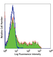
Human glioblastoma cell line LN319 stained with NC-08 PE -
Alexa Fluor® 647 anti-human Podoplanin

Human glioblastoma cell line LN319 stained with NC-08 Alexa ... -
Alexa Fluor® 488 anti-human Podoplanin

Human glioblastoma cell line LN 319 stained with NC-08 Alexa... -
PE/Cyanine7 anti-human Podoplanin

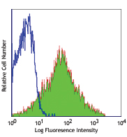
Human glioblastoma cell line LN319 was stained with podoplan... -
Biotin anti-human Podoplanin

Human glioblastoma cell line LN319 was stained with biotinyl... -
PerCP/Cyanine5.5 anti-human Podoplanin

Human glioblastoma cell line LN319 was stained with podoplan... -
Alexa Fluor® 594 anti-human Podoplanin

Human glioblastoma cell line, LN319 were fixed with 1% paraf... -
TotalSeq™-A0127 anti-human Podoplanin
-
APC/Cyanine7 anti-human Podoplanin

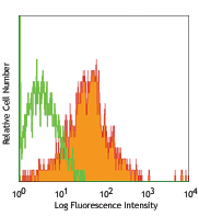
Human glioblastoma cell line LN319 was stained with podoplan... -
APC/Fire™ 750 anti-human Podoplanin

Human glioblastoma cell line LN319 was stained with podoplan... -
FITC anti-human Podoplanin

Human glioblastoma cell line LN319 was stained with podoplan... -
APC anti-human Podoplanin

Human glioblastoma cell line LN319 was stained with podoplan... -
PE/Dazzle™ 594 anti-human Podoplanin

Human glioblastoma cell line LN319 was stained with podoplan... -
TotalSeq™-C0127 anti-human Podoplanin
-
TotalSeq™-B0127 anti-human Podoplanin
-
TotalSeq™-D0127 anti-human Podoplanin


Follow Us