 Login/Register


- Clone
- 3A9 (See other available formats)
- Regulatory Status
- RUO
- Workshop
- HCDM listed
- Other Names
- PDCD6IP, PDCD6 interacting protein, AIP1, Hp95
- Isotype
- Mouse IgG1, κ
- Ave. Rating
- Submit a Review
- Product Citations
- publications

-

Total cell lysates (15 µg total protein) from HEK293T (WT) and HEK293T ALIX CRISPR/CAS9 knockout (KO) cells were resolved by 4-12% Bis-Tris gel electrophoresis, transferred to a nitrocellulose membrane, and probed with 2.0 µg/mL (1:250 dilution) of Purified anti-Alix Antibody, clone 3A9, overnight at 4°C. Proteins were visualized by chemiluminescence detection using HRP goat anti-mouse IgG Antibody (Cat. No. 405306) at a 1:3000 dilution. Direct-Blot™ HRP anti-β-Actin Antibody (Cat. No. 664804) was used as a loading control at a 1:25000 dilution (lower). Lane M: Molecular Weight marker. -

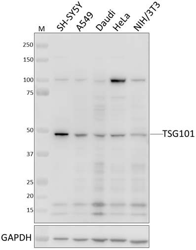
Whole cell extracts (15 µg total protein) from the indicated cell lines were resolved by 4-12% Bis-Tris gel electrophoresis, transferred to a PVDF membrane, and probed with 1 µg/mL of purified anti-Alix antibody (clone 3A9) overnight at 4°C. Proteins were visualized by chemiluminescence detection using HRP goat anti-mouse IgG antibody (Cat. No. 405306) at a 1:3000 dilution. Direct-Blot™ HRP anti-GAPDH antibody (Cat. No. 607904) was used as a loading control at a 1:50000 dilution (lower). Lane M: Molecular weight marker.
| Cat # | Size | Price | Quantity Check Availability | Save | ||
|---|---|---|---|---|---|---|
| 634501 | 25 µg | $112 | ||||
| 634502 | 100 µg | $226 | ||||
Alix (ALG-2-interacting protein X) is an adaptor protein that was first described for its capacity to bind to the calcium-binding protein ALG-2, the expression of which seemed necessary for cell death. The predicted molecular weight is approximately 96 kD. Alix contains an N-terminal Bro1 domain and a C-terminal proline-rich domain (PRD). The PRD contains multiple polyproline motifs, which are potential docking sites for proteins containing SH3 domains, interact with multiple cellular Alix-binding partners that are involved in apoptotic induction, endosomal sorting, and endocytosis. The Bro1 domain interacts with CHMP4b, which is also involved in endosomal sorting.
Product DetailsProduct Details
- Verified Reactivity
- Human, Mouse
- Reported Reactivity
- Rat, Frog
- Antibody Type
- Monoclonal
- Host Species
- Mouse
- Immunogen
- Recombinant full-length human Alix protein
- Formulation
- This antibody is provided in phosphate-buffered solution, pH 7.2, containing 0.09% sodium azide.
- Preparation
- The antibody was purified by affinity chromatography.
- Concentration
- 0.5 mg/ml
- Storage & Handling
- The antibody solution should be stored undiluted between 2°C and 8°C.
- Application
-
WB - Quality tested
ICC, IP - Reported in the literature, not verified in house - Recommended Usage
-
Each lot of this antibody is quality control tested by Western blotting. Western blotting, suggested working dilution(s): Use 1 µg/ml antibody dilution buffer for each mini-gel. It is recommended that the reagent be titrated for optimal performance for each application.
- Application Notes
-
Clone 3A9 has been shown to be useful for Western blotting1, immunoprecipitation, and immunofluorescence2 staining of the human and mouse Alix protein.
Clone 3A9 shows strong specificity for mouse Alix. -
Application References
(PubMed link indicates BioLegend citation) - Product Citations
-
- RRID
-
AB_2268110 (BioLegend Cat. No. 634501)
AB_2162471 (BioLegend Cat. No. 634502)
Antigen Details
- Structure
- Belongs to a conserved family of proteins that have in common an N-terminal Bro1 domain and a C-terminal proline-rich domain. Predicted molecular weight approximately 96 kD.
- Distribution
-
Cytoplasm. Alix is also abundant within extracellular vesicles known as exosomes, and enriched in virions that are released from cells under the direction of the HIV-1 Gag protein.
- Function
- Adaptor in signal transduction pathways. Involved in apoptosis and endosomal membrane trafficking and viral budding.
- Interaction
- ALG2, SH3GL2, H-Vps28, CHMP4B, CHMP4A, CHMP4C, SETA, CIN85, Src
- Biology Area
- Cell Biology
- Antigen References
-
1. Wu Y, et al. 2000. Differentiation 67:139.
2. Missotten M, et al. 1999. Cell Death Differ. 6:124.
3. Odorizzi G. 2006. J Cell Sci. 119:3025. - Gene ID
- 10015 View all products for this Gene ID
- UniProt
- View information about Alix on UniProt.org

Related FAQs
Other Formats
View All Alix Reagents Request Custom Conjugation| Description | Clone | Applications |
|---|---|---|
| Purified anti-Alix | 3A9 | WB,ICC,IP |
| Alexa Fluor® 594 anti-Alix | 3A9 | ICC |
Customers Also Purchased
Compare Data Across All Formats
This data display is provided for general comparisons between formats.
Your actual data may vary due to variations in samples, target cells, instruments and their settings, staining conditions, and other factors.
If you need assistance with selecting the best format contact our expert technical support team.
-
Purified anti-Alix

Total cell lysates (15 µg total protein) from HEK293T (WT) a... 
Whole cell extracts (15 µg total protein) from the indicated... -
Alexa Fluor® 594 anti-Alix

NIT/3T3 cells were fixed with -20°C Methanol for ten minutes...


Follow Us