 Login/Register


- Clone
- MIH6 (See other available formats)
- Regulatory Status
- RUO
- Other Names
- Programmed cell death ligand 1 (PD-L1), B7 homolog 1 (B7-H1), B7-H, B7H1, PDL1, PDCD1L1, PDCD1LG1
- Isotype
- Rat IgG2a, λ
- Ave. Rating
- Submit a Review
- Product Citations
- publications

-

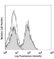
C57BL/6 mouse splenocytes were stained with LEAF™ purified CD274 (clone MIH6, filled histogram) or purified rat IgG2a, κ isotype control (open histogram), followed by anti-Rat IgG PE.
| Cat # | Size | Price | Quantity Check Availability | Save | ||
|---|---|---|---|---|---|---|
| 153602 | 100 µg | $149 | ||||
| 153603 | 1 mg | $512 | ||||
CD274, also known as B7-H1 or programmed death ligand 1 (PD-L1), is a 40 kD type I transmembrane protein and a member of the B7 family within the immunoglobulin receptor superfamily. It is expressed on T cells, B cells, NK cells, dendritic cells, IFN-γ activated endothelial cells, and monocytes. B7-H1 is one of the ligands of PD-1. The interaction of B7-H1 with PD-1 plays an important role in the inhibition of T cell responses. Other studies have shown that B7-H1 is able to costimulate T cell growth and cytokine production. CD274 is involved in costimulation essential for T cell proliferation and production of IL-10 and IFN-γ, in an IL-2-dependent and a PD-1-independent manner. Its interaction with PD-1 inhibits T cell proliferation and cytokine production.
Product DetailsProduct Details
- Verified Reactivity
- Mouse
- Antibody Type
- Monoclonal
- Host Species
- Rat
- Immunogen
- Mouse PD-L1-transfected cells
- Formulation
- 0.2 µm filtered in phosphate-buffered solution, pH 7.2, containing no preservative.
- Endotoxin Level
- Less than 0.01 EU/µg of the protein (< 0.001 ng/µg of the protein) as determined by the LAL test.
- Preparation
- The Ultra-LEAF™ (Low Endotoxin, Azide-Free) antibody was purified by affinity chromatography.
- Concentration
- The antibody is bottled at the concentration indicated on the vial, typically between 2 mg/mL and 3 mg/mL. Older lots may have also been bottled at 1 mg/mL. To obtain lot-specific concentration and expiration, please enter the lot number in our Certificate of Analysis online tool.
- Storage & Handling
- The antibody solution should be stored undiluted between 2°C and 8°C. This Ultra-LEAF™ solution contains no preservative; handle under aseptic conditions.
- Application
-
FC - Quality tested
Blocking - Reported in the literature, not verified in house - Recommended Usage
-
Each lot of this antibody is quality control tested by immunofluorescent staining with flow cytometric analysis. For flow cytometric staining, the suggested use of this reagent is ≤ 0.5 µg per million cells in 100 µl volume or 100 µl of whole blood. It is recommended that the reagent be titrated for optimal performance for each application.
- Application Notes
-
mAb MIH6 blocks the binding of mouse PD-L1 to PD-1 (CD279)
-
Application References
(PubMed link indicates BioLegend citation) -
- Gassner FJ, et al. 2015. Br J Haematol. 170:515 (Block)
- Haile ST, et al. 2013. J Immunol. 191:2829 (FC)
- Hirahara K, et al. 2012. Immunity. 36:1017 (Block)
- Fife BT, et al. 2009. Nat Immunol. 10:1185 (Block)
- Kanai T, et al. 2003. J Immunol. 171:4156 (Block)
- Product Citations
-
- RRID
-
AB_2715965 (BioLegend Cat. No. 153602)
AB_2715966 (BioLegend Cat. No. 153603)
Antigen Details
- Structure
- Type 1 transmembrane protein, member of the B7 family, 40kD.
- Distribution
-
T cells, B cells, NK cells, dendritic cells, IFN-γ activated endothelial cells, and monocytes
- Function
- CD274 is involved in the costimulatory signal, essential for T lymphocyte proliferation and production of IL-10 and IFN-γ, in an IL-2-dependent and a PD-1-CD1-independent manner. Its interaction with PD-1-CD1 inhibits T-cell proliferation and cytokine production.
- Ligand/Receptor
- PD-1 (CD279)
- Cell Type
- B cells, Dendritic cells, Endothelial cells, Monocytes, NK cells, T cells
- Biology Area
- Cancer Biomarkers, Costimulatory Molecules, Immunology
- Molecular Family
- Adhesion Molecules, CD Molecules, Immune Checkpoint Receptors
- Antigen References
-
1. Dorand RD. 2016. Science. 353:399.
2. Khan AR, et al. 2015. Nat Commun. 6:5997.
3. Kiyasu J, et al. 2015. Blood. 126:2193
4. Herold M, et al. 2015. J Immunol. 195:3584
5. Buddhisa S, et al. 2015. J Immunol. 194:4413 - Gene ID
- 60533 View all products for this Gene ID
- UniProt
- View information about CD274 on UniProt.org


Related FAQs
- Do you guarantee that your antibodies are totally pathogen free?
-
BioLegend does not test for pathogens in-house aside from the GoInVivo™ product line. However, upon request, this can be tested on a custom basis with an outside, independent laboratory.
- Does BioLegend test each Ultra-LEAF™ antibody by functional assay?
-
No, BioLegend does not test Ultra-LEAF™ antibodies by functional assays unless otherwise indicated. Due to the possible complexities and variations of uses of biofunctional antibodies in different assays and because of the large product portfolio, BioLegend does not currently perform functional assays as a routine QC for the antibodies. However, we do provide references in which the antibodies were used for functional assays and we do perform QC to verify the specificity and quality of the antibody based on our strict specification criteria.
- Does BioLegend test each Ultra-LEAF™ antibody for potential pathogens?
-
No, BioLegend does not test for pathogens in-house unless otherwise indicated. However, we can recommend an outside vendor to perform this testing as needed.
- Have you tested this Ultra-LEAF™ antibody for in vivo or in vitro applications?
-
We don't test our antibodies for in vivo or in vitro applications unless otherwise indicated. Depending on the product, the TDS may describe literature supporting usage of a particular product for bioassay. It may be best to further consult the literature to find clone specific information.
Other Formats
View All CD274 Reagents Request Custom Conjugation| Description | Clone | Applications |
|---|---|---|
| Ultra-LEAF™ Purified anti-mouse CD274 (B7-H1, PD-L1) | MIH6 | FC,Block |
| TotalSeq™-A0190 anti-mouse CD274 (B7-H1, PD-L1) | MIH6 | PG |
| Brilliant Violet 605™ anti-mouse CD274 (B7-H1, PD-L1) | MIH6 | FC |
| TotalSeq™-B0190 anti-mouse CD274 (B7-H1, PD-L1) | MIH6 | PG |
| TotalSeq™-C0190 anti-mouse CD274 (B7-H1, PD-L1) | MIH6 | PG |
| PE anti-mouse CD274 (B7-H1, PD-L1) | MIH6 | FC |
| PE/Cyanine7 anti-mouse CD274 (B7-H1, PD-L1) | MIH6 | FC |
| APC anti-mouse CD274 (B7-H1, PD-L1) | MIH6 | FC |
Customers Also Purchased
Compare Data Across All Formats
This data display is provided for general comparisons between formats.
Your actual data may vary due to variations in samples, target cells, instruments and their settings, staining conditions, and other factors.
If you need assistance with selecting the best format contact our expert technical support team.
-
Ultra-LEAF™ Purified anti-mouse CD274 (B7-H1, PD-L1)

C57BL/6 mouse splenocytes were stained with LEAF™ purified C... -
TotalSeq™-A0190 anti-mouse CD274 (B7-H1, PD-L1)
-
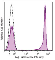
Brilliant Violet 605™ anti-mouse CD274 (B7-H1, PD-L1)

C57BL/6 mouse splenocytes were stained with CD274 (B7-H1, PD... -
TotalSeq™-B0190 anti-mouse CD274 (B7-H1, PD-L1)
-
TotalSeq™-C0190 anti-mouse CD274 (B7-H1, PD-L1)
-
PE anti-mouse CD274 (B7-H1, PD-L1)

C57BL/6 mouse splenocytes were stained with CD274 (B7-H1, PD... -
PE/Cyanine7 anti-mouse CD274 (B7-H1, PD-L1)

C57BL/6 mouse splenocytes were stained with anti-mouse CD274... -
APC anti-mouse CD274 (B7-H1, PD-L1)

C57 mouse splenocytes were stained with anti-mouse CD4 Brill...


Follow Us