 Login/Register


- Clone
- 12F7 (See other available formats)
- Regulatory Status
- RUO
- Other Names
- Catenin (Cadherin-Associated Protein), Beta 1, 88 kD, CTNNB1, TNNB, MRD19, armadillo, catenin beta-1
- Isotype
- Mouse IgG1, κ
- Ave. Rating
- Submit a Review
- Product Citations
- publications

-

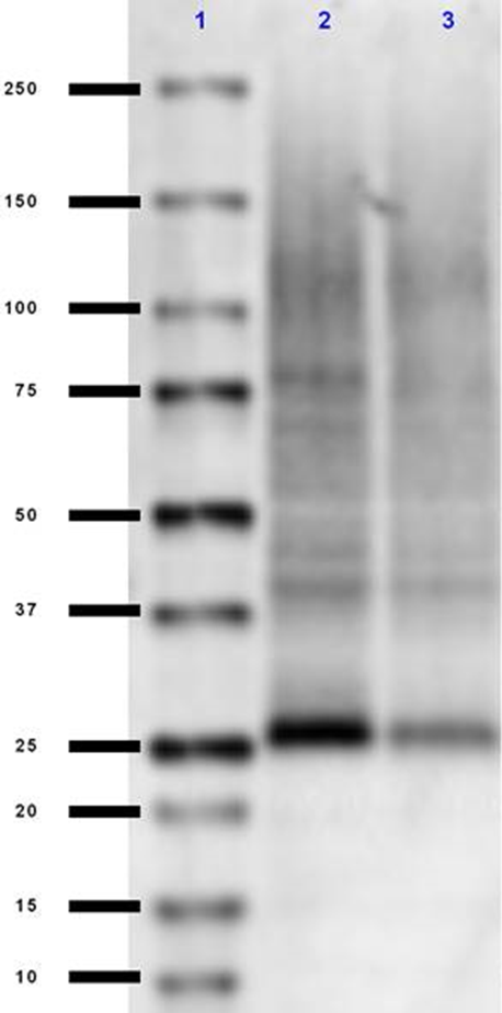
Western blot of purified anti-β Catenin 1 (CTNNB1) antibody (clone 12F7). Lane 1: Molecular weight marker; Lane 2: 20 µg of human brain lysate; Lane 3: 20 µg of mouse brain lysate; Lane 4: 20 µg of rat brain lysate. The blot was incubated with 1 µg/mL of the primary antibody overnight at 4°C, followed by incubation with HRP labeled goat anti-mouse IgG (Cat. No. 405306). Enhanced chemiluminescence was used as the detection system. -

Paraffin-embedded mouse testicle sections stained with anti-β Catenin 1 antibody. Heat-induced antigen retrieval was performed using pH 6.0 sodium citrate in a pressure cooker. Blocking was performed using Mouse-on-Mouse blocking reagents. The section was stained with 10 µg/mL of anti-β Catenin 1 (clone 12F7) for 30 minutes and color developed using DAB. Nuclei were counterstained with Nuclear Fast Red for two minutes. This image was captured under a 40X objective. Credit: Hans Snyder, Histologics. -

Human colon adenocarcinoma cell line SW480 treated (open historgram) or untreated (filled histogram) with tankyrase inhibitor XAV939 for 18 hours, then treated with True-Nuclear™ Transcription FactorBuffer set (Cat. No. 424401) and stained with purified anti-β Catenin 1 (clone 12F7), followed by anti-mouse IgG PE.
| Cat # | Size | Price | Quantity Check Availability | Save | ||
|---|---|---|---|---|---|---|
| 844601 | 25 µg | $112 | ||||
| 844602 | 100 µg | $288 | ||||
β-catenin is part of a complex of proteins that constitute adherens junctions (AJs). AJs are necessary for the creation and maintenance of epithelial cell layers by regulating cell growth and adhesion between cells. The encoded protein also anchors the actin cytoskeleton and may be responsible for transmitting the contact inhibition signal that causes cells to stop dividing once the epithelial sheet is complete. β-catenin also plays a key role in Wnt signaling pathways and thus is involved in neural differentiation, synaptic plasticity, neurodegenerative disease, and prevention of apoptosis. Finally, this protein binds to the product of the adenomatous polyposis coli (APC) gene, which is mutated in the adenomatous polyposis of the colon. Mutations in this gene are a cause of colorectal cancer (CRC), pilomatrixoma (PTR), medulloblastoma (MDB), and ovarian cancer.
Product DetailsProduct Details
- Verified Reactivity
- Human, Mouse, Rat
- Antibody Type
- Monoclonal
- Host Species
- Mouse
- Immunogen
- Recombinant β-catenin (N-terminal fragment) fused to MBP.
- Formulation
- Phosphate-buffered solution, pH 7.2, containing 0.09% sodium azide.
- Preparation
- The antibody was purified by affinity chromatography.
- Concentration
- 0.5 mg/ml
- Storage & Handling
- The antibody solution should be stored undiluted between 2°C and 8°C.
- Application
-
WB - Quality tested
IHC-P, ICFC - Verified - Recommended Usage
-
Each lot of this antibody is quality control tested by Western blotting. For Western blotting, the suggested use of this reagent is 1.0 - 10 µg/mL. For immunohistochemistry on formalin-fixed paraffin-embedded tissue sections, a concentration of 10 µg/mL is suggested. For intracellular flow cytometry using our True-Nuclear™ Transcription Factor Staining Protocol, the suggested use of this reagent is ≤ 0.25 µg per million cells in 100 µL volume. It is recommended that the reagent be titrated for optimal performance for each application.
- Application Notes
-
Additional reported applications (for the relevant formats) include: immunfluorescence microscopy4 and immunohistochemical staining3.
For in vivo studies or highly sensitive assays, we recommend Ultra-LEAF™ purified antibody (Cat. No. 844603) with a lower endotoxin limit than standard LEAF™ purified antibodies (Endotoxin <0.01 EU/µg). -
Application References
(PubMed link indicates BioLegend citation) -
- Sacco P, et al. 1995. J. Biol. Chem. 270:20201. (WB)
- Johnson KR, et al. 1993. Exp. Cell Res. 207:252.
- Gupta K, et al. 2012. J. Ped. Hem. Onc. 34:320. (IHC-P)
- Radice G, et al. 1997. Dev. Bio. 181:64. (IHC-P)
- Product Citations
-
- RRID
-
AB_2750041 (BioLegend Cat. No. 844601)
AB_2565803 (BioLegend Cat. No. 844602)
Antigen Details
- Structure
- 88 kD armadillo repeat containing protein with an unstructured N and C terminal.
- Distribution
-
β-catenin localizes to cell-cell contacts (adherens junctions) and the cell membrane as well as the cytoplasmic pool of protein. β-catenin can also translocate to the nucleus and is in interphase found in puncta at the proximal end of centrioles. In mitosis, it localizes to the centrosomes at the spindle.
- Function
- Wnt signaling pathway, regulation of cell-cell adhesion, neurogenesis, transcription, transcription regulation, early embryonic patterning, asymmetric cell division, stem cell renewal, and the epithelial to mesenchymal transition.
- Interaction
- Wnt signaling pathway, apoptotic cleavage of cellular proteins, and EphB-EphrinB signaling.
- Biology Area
- Cell Adhesion, Cell Biology, Neuroscience, Signal Transduction, Synaptic Biology
- Molecular Family
- Adhesion Molecules
- Antigen References
-
1. Hirano S, et al. 2012. Physiol. Rev. 92:597.
- Gene ID
- 1499 View all products for this Gene ID
- UniProt
- View information about beta Catenin 1 on UniProt.org

Related FAQs
Other Formats
View All β Catenin 1 Reagents Request Custom Conjugation| Description | Clone | Applications |
|---|---|---|
| Purified anti-β Catenin 1 (CTNNB1) | 12F7 | WB,IHC-P,ICFC |
| Ultra-LEAF™ Purified anti-β Catenin 1 (CTNNB1) | 12F7 | WB |
| PE anti-β Catenin 1 (CTNNB1) | 12F7 | ICFC |
| APC anti-β Catenin 1 (CTNNB1) | 12F7 | ICFC |
Customers Also Purchased
Compare Data Across All Formats
This data display is provided for general comparisons between formats.
Your actual data may vary due to variations in samples, target cells, instruments and their settings, staining conditions, and other factors.
If you need assistance with selecting the best format contact our expert technical support team.
-
Purified anti-β Catenin 1 (CTNNB1)

Western blot of purified anti-β Catenin 1 (CTNNB1) antibody ... 
Paraffin-embedded mouse testicle sections stained with anti-... 
Human colon adenocarcinoma cell line SW480 treated (open his... -
Ultra-LEAF™ Purified anti-β Catenin 1 (CTNNB1)

Western blot was incubated with either clone 12F7 (lanes 2-3... -
PE anti-β Catenin 1 (CTNNB1)

Human colon adenocarcinoma cell line SW480 treated (open his... -
APC anti-β Catenin 1 (CTNNB1)

SW480 cells (Human colon adenocarcinoma cell line) were trea...


Follow Us