- Regulatory Status
- RUO
- Ave. Rating
- Submit a Review
- Product Citations
- publications

-

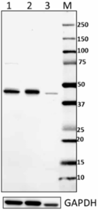
Total lysates (15 µg protein) from HeLa (lane 1), Molt-4 (lane 2) and NIH3T3 (lane 3) were resolved by electrophoresis (4-12% Bis-Tris gel), transferred to nitrocellulose, and probed with 1:500 diluted (1 µg/mL) purified anti-IRF1 antibody (upper). Proteins were visualized using an HRP goat anti-mouse-IgG secondary antibody (cat. No. 405306) or HRP Donkey anti-rabbit IgG Antibody (Cat. No. 406401) for GAPDH and chemiluminescence detection. GAPDH (poly6314, Cat. No. 631401) antibody was used as a loading control (lower). Lane M: MW ladder -

Total lysates (15 µg protein) from HeLa (lane 1) were resolved by electrophoresis (4-12% Bis-Tris gel), transferred to nitrocellulose, and probed with 1:500 diluted (1 µg/mL) purified anti-IRF2 antibody (upper). Proteins were visualized using an HRP goat anti-mouse-IgG secondary antibody (Cat. No. 405306). Direct-Blot™ HRP anti-β-actin Antibody (Cat. No. 643807) was used as a loading control (lower). Lane M: MW ladder. -

Total lysates (15 µg protein) from Daudi (lane 1) and A20 (lane 2) were resolved by electrophoresis (4-12% Bis-Tris gel), transferred to nitrocellulose, and probed with 1:1000 diluted (0.5 µg/mL) purified anti-IRF3 antibody (upper). Proteins were visualized using an HRP goat anti-mouse-IgG secondary antibody (Cat. No. 405306) or HRP Donkey anti-rabbit IgG Antibody (Cat. No. 406401) for GAPDH and chemiluminescence detection. GAPDH (poly6314, Cat. No. 631401) antibody was used as a loading control (lower). Lane M: MW ladder. -

Total lysates (15 µg protein) from Daudi (lane 1) and A20 (lane 2) were resolved by electrophoresis (4-12% Bis-Tris gel), transferred to nitrocellulose, and probed with 1:1000 diluted (0.5 µg/mL) purified anti-IRF4 antibody (upper). Proteins were visualized using an HRP goat anti-rat-IgG secondary antibody (Cat. No. 405405) or HRP Donkey anti-rabbit IgG Antibody (Cat. No. 406401) for GAPDH and chemiluminescence detection. GAPDH (poly6314, Cat. No. 631401) antibody was used as a loading control (lower). Lane M: MW ladder. -

Total lysates (15 µg protein) from 293E (lane 1), THP-1 (lane 2) and Raw264.7 (lane 3) were resolved by electrophoresis (4-12% Bis-Tris gel), transferred to nitrocellulose, and probed with 1:2500 diluted (0.2 µg/mL) purified anti-IRF5 antibody (upper). Proteins were visualized using an HRP goat anti-mouse-IgG secondary antibody (Cat. No. 405306) or HRP Donkey anti-rabbit IgG Antibody (Cat. No. 406401) for GAPDH and chemiluminescence detection. GAPDH (poly6314, Cat. No. 631401) antibody was used as a loading control (lower). Lane M: MW ladder. -

Total lysates (15 µg protein) from Jurkat (lane 1), HaCaT (lane 2) and NIH3T3 (lane 3) were resolved by electrophoresis (4-12% Bis-Tris gel), transferred to nitrocellulose, and probed with 1:1000 diluted (0.5 µg/mL) purified anti-IRF6 antibody (upper). Proteins were visualized using an HRP goat anti-mouse-IgG secondary antibody (Cat. No. 405306) or HRP Donkey anti-rabbit IgG Antibody (Cat. No. 406401) for GAPDH and chemiluminescence detection. GAPDH (poly6314, Cat. No. 631401) antibody was used as a loading control (lower). Lane M: MW ladder.
| Cat # | Size | Price | Quantity Check Availability | Save | ||
|---|---|---|---|---|---|---|
Interferon regulatory transcription factor (IRF) family proteins play important roles in mediating immune response and regulating cell cycle progression, tumor suppression, and apoptosis. IRF1 is the first identified member. Its expression is induced in response to pathogen infection or the stimulation of several cytokines. IRF2 is mainly functioning as a transcriptional repressor. It binds to the interferon-sensitive response elements (ISREs) and competes for binding sites with the other IRF transcription factors, such as IRF1 and IRF9. IRF3 mediates ISRE promoter activation and functions as a molecular switch for antiviral activity. IRF4 and IRF8 are highly homologous to each other and also redundant in function. IRF4 is critical for Th2 and Th17 development. IRF-5 participates in the virus-mediated activation of interferon and regulates gene expression induced by cell growth, differentiation, and apoptosis. IRF6 is not involved in the expression of interferon genes and doesn’t seem to be involved in innate immunity. Data suggests that IRF6 is necessary for appropriate epidermal differentiation and development. Upon virus infection, IRF-7 forms a complex with MyD88 and TRAF6 when stimulated by toll-like receptors TLR7, TLR8, and TLR9, which in turn results in nuclear translocation and induction of interferon gene expression. IRF8 regulates hematopoietic cell growth and differentiation of macrophage and dendritic cells. The activated STAT1:STAT2 heterodimer recruits IRF9 and forms a heterotrimeric complex termed IFN-stimulated gene factor 3 (ISGF3). The ISGF3 complex, in turn, translocates to the nucleus and transactivates IFN-inducible genes.
Product DetailsKit Contents
- Kit Contents
-
Specificity Clone Size Reactivity Isotype Pred. MW (kD) Anti-IRF1 13H3A44 100 μg Human Mouse IgG2a, κ 37 kD Anti-IRF2 13B2A38 100 μg Human, Mouse Mouse IgG1, κ 39 kD Anti-IRF3 12A4A35 100 μg Human, Mouse Mouse IgG2b, κ 47.2 kD Anti-IRF4 IRF4.3E4 100 μg Mouse, Human Rat IgG1, κ 51 kD Anti-IRF5 11F4A09 100 μg Human Mouse IgG2b, κ 56 kD Anti-IRF6 14B2C16 100 μg Human Mouse IgG2a, κ 53 kD Anti-IRF7 12G9A36 100 μg Human Mouse IgG2b, κ 54 kD Anti-IRF8 7G11A45 100 μg Human Mouse IgG1, κ 48 kD Anti-IRF9 5A3A39 100 μg Human Mouse IgG2b, κ 44 kD * For detailed information about each specificity, please follow the provided links.
Product Details
- Formulation
- Please refer to individual product datasheets for details.
- Storage & Handling
- Upon receipt, store undiluted at 2-8°C.
- Recommended Usage
-
Each lot of antibodies in this kit is quality control tested by Western Blotting. For Western blotting, the suggested uses of these reagents are as follows:
Anti-IRF1: 0.5 - 2.0 µg/ml (1:250 - 1:1000 dilution)
Anti-IRF2: 0.5 - 2.0 µg/ml (1:250 - 1:1000 dilution)
Anti-IRF3: 0.5 - 2.0 µg/ml (1:250 - 1:1000 dilution)
Anti-IRF4: 0.1 - 1.0 µg/ml (1:500 - 1:5000 dilution)
Anti-IRF5: 0.2 - 1.0 µg/ml (1:500 - 1:2500 dilution)
Anti-IRF6: 0.2 - 2.5 µg/ml (1:200 - 1:1000 diltuion)
Anti-IRF7: 0.2 - 2.0 µg/ml (1:250 - 1:2500 dilution)
Anti-IRF8: 0.5 - 2.0 µg/ml (1:250 - 1:1000 dilution)
Anti-IRF9: 0.5 - 2.5 µg/ml (1:500 - 1:5000 dilution)
It is recommended that the reagent be titrated for optimal performance for each application. - Application Notes
-
For verified or reported applications for these antibodies, please see individual product datasheets.
Antigen Details
- Biology Area
- Cell Biology, Immunology, Innate Immunity, Transcription Factors
- Molecular Family
- Nuclear Markers
- Antigen References
-
1. Frontini M, et al. 2009. Nucleic Acids Res. 37:1073.
2. Yarilina A, et al. 2008. Nat. Immunol. 9:378.
3. Hayashi H, et al. 2011. Proc. Natl. Acad. Sci. USA. 108:18766.
4. Hida S, et al. 2005. Blood. 106:2011.
5. Lin R, et al. 1999. Mol. Cell Biol. 19:959.
6. Hiscott J, et al. 2007. J. Biol. Chem. 282:15325.
7. Lu R. 2008. Trends Immunol 29:487.
8. Barnes BJ, et al. 2003. J. Biol. Chem. 278:16630.
9. Rullo OJ, et al. 2010. Ann. Rheum. Dis. 69:611.
10. Botti E, et al. 2011. Proc. Natl. Acad. Sci. USA 108:13710.
11. Restivo G, et al. 2011. EMBO 30:4571.
12. Yu Y, et al. 2010. Immunity. 33:863.
13. Liang Q, et al. 2011. J. Immunol. 186:1001.
14. Ouyang X, et al. 2011. Nat. Commun. 2:314.
15. Thibault DL, et al. 2008. J. Clin. Invest. 118:1417.
16. Kraus TA, et al. 2003. J. Biol. Chem. 278:13033.
17. Xiao W, et al. 2001. J. Biol. Chem. 276:23275. - Gene ID
- 3659 View all products for this Gene ID 3660 View all products for this Gene ID 3661 View all products for this Gene ID 3662 View all products for this Gene ID 3663 View all products for this Gene ID 3664 View all products for this Gene ID 3665 View all products for this Gene ID 3394 View all products for this Gene ID 10379 View all products for this Gene ID
- App Abbreviation (DOES NOT SHOW ON TDS):
- WB
 Login/Register


Follow Us