


- Clone
- A17020B (See other available formats)
- Regulatory Status
- RUO
- Other Names
- S6, 40S ribosomal protein S6
- Isotype
- Mouse IgG1, κ
- Ave. Rating
- Submit a Review
- Product Citations
- publications

-

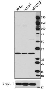
Total cell lysates (15 µg protein) from serum-starved Jurkat cells treated without (-) or with (+) 160 nM PMA for 15 minutes were resolved by 4-12% Bis-Tris gel electrophoresis, transferred to nitrocellulose, and probed with the 1.0 µg/mL (1:500 dilution) of purified anti-RPS6 Phospho (Ser235/Ser236) antibody (Clone A17020B). Proteins were visualized by chemiluminescence detection using HRP goat anti-mouse-IgG (Cat. No. 405301) at a 1:3000 dilution. Equal protein loading was confirmed using a pan RPS6 antibody. Lane M: MW ladder. -

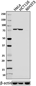
Total cell lysates (15 µg protein) from serum-starved NIH/3T3 cells treated without (-) or with 100 ng/mL PDGF-BB (Cat. No. 558804) for 20 minutes were resolved by 4-12% Bis-Tris gel electrophoresis, transferred to nitrocellulose, and probed with the 1.0 µg/mL (1:500 dilution) of purified anti-RPS6 Phospho (Ser235/Ser236) antibody (Clone A17020B). Proteins were visualized by chemiluminescence detection using HRP goat anti-mouse-IgG (Cat. No. 405301) at a 1:3000 dilution. Equal protein loading was confirmed using a pan RPS6 antibody. Lane M: MW ladder. -

Serum starved NIH/3T3 cells were untreated (panel A) or stimulated with 100 ng/mL PDGF-BB for 20 minutes. Cell were then fixed with 4% paraformaldehyde for 10 minutes, permeabilized with 0.5% Triton X-100 for 10 minutes, and blocked with 5% FBS for 60 minutes. Cells were then intracellularly stained with 1:100 (5 µg/mL) of purified anti-RPS6 Phospho (Ser235/Ser236) antibody overnight at 4°C, after which proteins were visualized with Alexa Fluor® 594 goat anti-mouse IgG (Cat. No. 405308) at 2.0 µg/mL. Nuclei were counterstained with DAPI, and the image was captured with a 60X objective. -

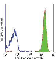
Human peripheral blood lymphocytes were stimulated with (filled histogram) or without (open histogram) Cell Activation cocktail without Brefeldin A (Cat. No. 423302) for 15 minutes, fixed with Fixation Buffer, permeabilied with True-Phos™ Perm Buffer (Cat. No. 425401), and intracellularly stained with purified anti-RPS6 Phosopho (Ser235/Ser236) (clone A17020B), followed by anti-mouse IgG PE. -

IHC staining of purified anti-RPS6 (Ser235/Ser236) (clone A17020B) on formalin-fixed paraffin-embedded human thymus. Following antigen retrieval using 1X Tris-EDTA, pH 9.0, the tissue was treated with (+) or without (-) Lambda Protein Phosphatase overnight, blocked, and incubated with 10.0 µg/mL purified anti-RPS6 (Ser235/Ser236) (Clone A17020B) (panel A) or 5.0 µg/mL of the anti-RPS6 (Cat. 691802) (panel B) overnight at 4°C. This was followed by incubation with Alexa Fluor® 647 goat anti-mouse IgG (Cat. No. 405322) at 2.5 µg/mL. Nuclei were counterstained with DAPI (blue) (Cat. No. 422801) and the slide was mounted with ProLong™ Gold Antifade Mountant. The image was captured with a 40X objective. Scale: 50 µm -

IHC staining of purified anti-RPS6 (Ser235/Ser236) (clone A17020B) on formalin-fixed paraffin-embedded human thymus without DAPI. The image was captured with a 40X objective. Scale: 50 µm.
| Cat # | Size | Price | Save |
|---|---|---|---|
| 608601 | 25 µg | ¥24,640 | |
| 608602 | 100 µg | ¥58,300 |
Ribosomal protein S6 (RPS6) is a key component of the small 40S ribosomal subunit and is the major substrate of protein kinases in eukaryotic ribosomes. In response to various cellular stimuli such as mitogenic stimulation, insulin, and increased nutrient availability, upstream kinases such as RSK and p70 kinases phosphorylate RPS6 at multiple serine sites. These modifications facilitate the recruitment of the 7-methylguanasine cap complex, thereby promoting the assembly of the translational pre-initiation complex and increased cellular protein synthesis capacity. RPS6 has been shown to be hyperphosphorylated in certain cancers, and phosphorylation is a critical determinant of pancreatic β–cell size and systemic glucose homeostasis function in diabeteic mouse models.
Product DetailsProduct Details
- Verified Reactivity
- Human, Mouse
- Antibody Type
- Monoclonal
- Host Species
- Mouse
- Immunogen
- Synthetic peptide from human RPS6 phosphorylated at Serines 235 and 236
- Formulation
- Phosphate-buffered solution, pH 7.2, containing 0.09% sodium azide.
- Preparation
- The antibody was purified by affinity chromatography.
- Concentration
- 0.5 mg/ml
- Storage & Handling
- The antibody solution should be stored undiluted between 2°C and 8°C.
- Application
-
WB - Quality tested
ICC, ICFC, IHC-P - Verified - Recommended Usage
-
Each lot of this antibody is quality control tested by Western blotting. For Western blotting, the suggested use of this reagent is 0.1 - 1.0 µg per ml. For immunocytochemistry, a concentration range of 1.0 - 5.0 μg/ml is recommended. For intracellular flow cytometry using our True-Phos™ Perm Buffer in Cell Suspension, the suggested use of this reagent is ≤ 0.25 µg per million cells in 100 µl volume. For immunohistochemistry on formalin-fixed paraffin-embedded tissue sections, a concentration of 10.0 µg/mL is suggested.It is recommended that the reagent be titrated for optimal performance for each application.
- Application Notes
-
Due to complete conservation of the immunizing sequence between humans, mouse and rat, this clone is is predicted to react with rat RPS6 phosphorylated at serines 235 and 236.
- RRID
-
AB_2749900 (BioLegend Cat. No. 608601)
AB_2749899 (BioLegend Cat. No. 608602)
Antigen Details
- Structure
- RPS6 is a 249 amino acid protein with a predicted molecular weight of 28.6 kD.
- Distribution
-
Cytosol, nucleus, ER, ubiquitous expression.
- Function
- Phosphorylation events at a cluster of serine residues (Ser 235, 236, 240, and 244) at the carboxy-terminus of RPS6 results in increased global translational efficiency. These sites are dephosphorylated during growth arrest.
- Interaction
- RPS6AK1 and RPS6AK3; other subunits of 40S ribosomal complex.
- Biology Area
- Cell Biology, Cell Proliferation and Viability, Protein Synthesis, Signal Transduction
- Molecular Family
- Nuclear Markers, Phospho-Proteins
- Antigen References
-
- Jefferies HB, et al. 1997. EMBO J. 16:3693.
- Ruvinsky I, et al. 2005. Genes. Dev.19:2199.
- Chumacher AM, et al. 2006. Biochemistry. 45:13614
- Roux PP, et al. 2007. J. Biol. Chem. 282:14056.
- Stevens C, et al. 2009. J. Biol. Chem. 284:334.
- Schlafli P, et al. 2011. FEBS J. 278:1757.
- Gene ID
- 6194 View all products for this Gene ID
- UniProt
- View information about RPS6 Phospho Ser235/Ser236 on UniProt.org

Related FAQs
Other Formats
View All RPS6 Phospho (Ser235/Ser236) Reagents Request Custom Conjugation| Description | Clone | Applications |
|---|---|---|
| Purified anti-RPS6 Phospho (Ser235/Ser236) | A17020B | WB,ICC,ICFC,IHC-P |
| PE anti-RPS6 Phospho (Ser235/Ser236) | A17020B | ICFC |
| PE/Cyanine7 anti-RPS6 Phospho (Ser235/Ser236) | A17020B | ICFC |
| PerCP/Cyanine5.5 anti-RPS6 Phospho (Ser235/Ser236) | A17020B | ICFC |
| Brilliant Violet 421™ anti-RPS6 Phospho (Ser235/Ser236) | A17020B | ICFC |
| Direct-Blot™ HRP anti-RPS6 Phospho (Ser235/Ser236) | A17020B | WB |
Customers Also Purchased
Compare Data Across All Formats
This data display is provided for general comparisons between formats.
Your actual data may vary due to variations in samples, target cells, instruments and their settings, staining conditions, and other factors.
If you need assistance with selecting the best format contact our expert technical support team.
-
Purified anti-RPS6 Phospho (Ser235/Ser236)

Total cell lysates (15 µg protein) from serum-starved Jurkat... 
Total cell lysates (15 µg protein) from serum-starved NIH/3T... 
Serum starved NIH/3T3 cells were untreated (panel A) or stim... 
Human peripheral blood lymphocytes were stimulated with (fil... 
IHC staining of purified anti-RPS6 (Ser235/Ser236) (clone A1... 
IHC staining of purified anti-RPS6 (Ser235/Ser236) (clone A1... -
PE anti-RPS6 Phospho (Ser235/Ser236)

Human peripheral blood lymphocytes were stimulated with (fil... -
PE/Cyanine7 anti-RPS6 Phospho (Ser235/Ser236)

Human peripheral blood lymphocytes were stimulated with (fil... -
PerCP/Cyanine5.5 anti-RPS6 Phospho (Ser235/Ser236)

Human peripheral blood lymphocytes were stimulated with (fil... -
Brilliant Violet 421™ anti-RPS6 Phospho (Ser235/Ser236)

Human peripheral blood lymphocytes were stimulated with (fil... -
Direct-Blot™ HRP anti-RPS6 Phospho (Ser235/Ser236)

Total cell lysates (15 µg protein) from serum-starved NIH/3T...


Follow Us