


- Clone
- W17241A (See other available formats)
- Regulatory Status
- RUO
- Other Names
- PIEZO Type Mechanosensitive Ion Channel Component 1, Membrane Protein Induced By Beta-Amyloid Treatment, FAM38A, Mib, KIAA023, LMPH3, DHS
- Isotype
- Rat IgG2b, κ
- Ave. Rating
- Submit a Review
- Product Citations
- publications

-

IHC staining of purified anti-PIEZO1 (clone W17241A) on human bladder. Tissue was fixed with Fixation Buffer (Cat. No. 420801), blocked with 5% FBS + 5% Normal Blocking Serum (Cat. No. 927503), and incubated with 10.0 µg/mL purified rat IgG2b, κ isotype control (Cat. No. 400602) (panel A) or 10.0 µg/mL of anti-PIEZO1 antibody (panel B) overnight at 4°C. This was followed by incubation with Alexa Fluor® 647 Goat anti-rat IgG (Cat. No. 405416) at 2.5 µg/mL. Nuclei were counterstained with DAPI (blue) and the slide was mounted with ProLong™ Gold Antifade Mountant. The image was captured with a 40X objective. Scale: 50 µm -

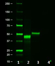
Whole cell lysates from HeLa (15 μg) untreated or treated with non-targeting siRNA (siCON) or siRNA targeting PIEZO1 (siPIEZO1) were resolved by 4-12% Bis-Tris gel electrophoresis, transferred to a PVDF membrane and probed with 1.0 μg/mL (1:500 dilution) of purified anti-PIEZO1 (clone W17241A) overnight at 4°C. Proteins were visualized by chemiluminescence detection using HRP goat anti-rat IgG (Cat. No. 405405) at a 1:3000 dilution. Direct-Blot™ HRP anti-GAPDH (Cat. No. 607904) was used as a loading control at a 1:50000 dilution (lower). Lane M: Molecular weight marker -

Whole cell extracts (15 µg total protein) from HeLa, A-431, and PC-3 cells were heated to 37°C for 30 minutes and resolved by 4-12% Bis-Tris gel electrophoresis, transferred to a PVDF membrane, and probed with 1.0 μg/mL (1:500 dilution) purified anti-PIEZO1 (clone W17241A) overnight at 4°C. Proteins were visualized by chemiluminescence detection using HRP goat anti-rat IgG (Cat. No. 405405) at a 1:3000 dilution. Western-Ready™ ECL Substrate Premium Kit (Cat. No. 426319) was used as a detection agent. Direct-Blot™ HRP anti-β-actin (Cat. No. 643807) was used as a loading control at a 1:10000 dilution (lower). Lane M: Molecular weight marker
| Cat # | Size | Price | Save |
|---|---|---|---|
| 602751 | 25 µg | ¥25,960 | |
| 602752 | 100 µg | ¥64,460 |
PIEZO1 and its paralog PIEZO2 are members of a non-selective cationic mechanosensitive channel family first discovered in a neuronal cell line through mechanical stimulation. It responds to mechanical forces such as static pressure, shear stress, and membrane stretch events by allowing the efflux of calcium ions from the ER lumen into the cytosol. The increase in cytosolic calcium results in phosphorylation of ERK1/2 and a commensurate induction of downstream mitotic genes, and can also promote prostate cancer development via activation of the Akt/mTOR signaling axis. PIEZO1 is overexpressed in many tumors and is a key regulator of tumor stiffness.
Product DetailsProduct Details
- Verified Reactivity
- Human, Mouse
- Antibody Type
- Monoclonal
- Host Species
- Rat
- Immunogen
- Partial recombinant human PIEZO1 protein
- Formulation
- Phosphate-buffered solution, pH 7.2, containing 0.09% sodium azide
- Preparation
- The antibody was purified by affinity chromatography.
- Concentration
- 0.5 mg/mL
- Storage & Handling
- The antibody solution should be stored undiluted between 2°C and 8°C.
- Application
-
IHC-P - Quality tested
WB - Verified - Recommended Usage
-
Each lot of this antibody is quality control tested by formalin-fixed paraffin-embedded immunohistochemical staining. For immunohistochemistry, a concentration range of 1.0 - 10.0 µg/mL is suggested. For western blotting, the suggested use of this reagent is 1.0 µg/mL. It is recommended that the reagent be titrated for optimal performance for each application.
- Application Notes
-
When testing this clone for western blot, we observed a considerable loss of signal with lysates that were boiled in sample buffer compared to samples that were heated to 37°C. This is likely due to high temperatures causing aggregation of PIEZO1, which is a multi-pass transmembrane domain protein.
This clone was tested for immunocytochemistry on HeLa cells fixed with 4% PFA and permeabilized with either methanol or Triton X-100. Both methods resulted in high non-specific staining. - RRID
-
AB_2910478 (BioLegend Cat. No. 602751)
AB_2910478 (BioLegend Cat. No. 602752)
Antigen Details
- Structure
- PIEZO1 is a 2521 amino acid protein with a predicted molecular weight of 287 kD.
- Distribution
-
Ubiquitously expressed/Endoplasmic reticulum
- Function
- Mechanically-activated ion channel
- Cell Type
- Epithelial cells
- Biology Area
- Neuroscience, Signal Transduction
- Molecular Family
- Ion Channels
- Antigen References
-
- Gudipaty SA, et al. 2017. Nature. 543:118-121.
- Nonomura K, et al. 2018. PNAS. 115:12817-12822.
- Romac JM, et al. 2018. Nat Comm. 9:1715.
- Zhao C, et al. 2019. Front Endocrinol. 10:373.
- Williams E. 2019. Sci Signal. 12:598.
- Han Y, et al. 2019. Int J Onc. 55:629-644.
- Gene ID
- 9780 View all products for this Gene ID
- UniProt
- View information about PIEZO1 on UniProt.org

Related FAQs
Other Formats
View All PIEZO1 Reagents Request Custom Conjugation| Description | Clone | Applications |
|---|---|---|
| Purified anti-PIEZO1 | W17241A | IHC-P,WB |
Customers Also Purchased

Compare Data Across All Formats
This data display is provided for general comparisons between formats.
Your actual data may vary due to variations in samples, target cells, instruments and their settings, staining conditions, and other factors.
If you need assistance with selecting the best format contact our expert technical support team.


Follow Us