


- Clone
- W15110A (See other available formats)
- Regulatory Status
- RUO
- Other Names
- Interleukin-2-inducible T-cell kinase, T-cell-specific kinase, EMT, LYK, PSCTK2
- Isotype
- Mouse IgG1, κ
- Ave. Rating
- Submit a Review
- Product Citations
- publications

-

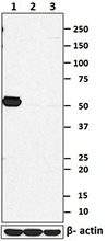
Total cell lysates (15 µg protein) from Jurkat (lane 1), HeLa (lane 2) and EL4 cells (lane 3) were resolved by electrophoresis (4-12% Bis-Tris gel), transferred to nitrocellulose, and probed with 1 µg/mL (1:500 dilution) of purified anti-ITK antibody, clone W15110A (upper panel). Proteins were visualized by chemiluminescence detection (Cat. No. 426303) using 1:3000 diluted anti-mouse-IgG secondary antibody conjugated to HRP for purified anti-ITK antibody or 1:10000 diluted Direct-Blot™ HRP anti-β-actin antibody, clone W16197A (lower panel). Lane M: Molecular weight ladder. -

Jurkat cells were fixed with 4% paraformaldehyde (PFA) for 15 minutes, permeabilized with 0.5% Triton X-100 for three minutes, and blocked with 1% BSA for 60 minutes. Then the cells were intracellularly stained with 5.0 µg/ml purified anti-ITK (clone W15110A) antibody for one hour, followed by staining with Alexa Fluor® 594 conjugated anti-mouse IgG antibody (red) and DAPI (blue) for one hour at room temperature. The image was captured with a 60X objective. -

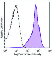
Human peripheral blood mononuclear cells were surface stained with CD3 Alexa Flour® 647 and then treated with True-Nuclear™ Transcription Factor Buffer Set. Cells were then stained with purified anti-ITK (clone W15110A) (left) or purified mouse IgG1, κ isotype (right). Followed by anti-mouse IgG1 PE. -

IL2-inducible T-cell kinase (ITK) is an intracellular tyrosine kinase that is expressed in T cells. It regulates the proliferation, differentiation and development of conventional T cells and NK T cells. This protein has 620 amino acids and contains one PH domain for cell membrane recruitment, one protein kinase domain, and both SH2 and SH3 domains which are commonly found in intracellular kinases. Once T cells are activated, a series of phosphorylation recruits and activates ITK through autophosphorylation. The activated ITK can activate downstream intracellular calcium mobilization and NFAT nucleus translocation, and in turn, activate T cells. The phenotypes of mice deficient in ITK show severely affected immune response and development.
Product DetailsProduct Details
- Verified Reactivity
- Human
- Antibody Type
- Monoclonal
- Host Species
- Mouse
- Immunogen
- Partial human ITK recombinant protein (1-200 a.a.) expressed in E. coli.
- Formulation
- Phosphate-buffered solution, pH 7.2, containing 0.09% sodium azide.
- Preparation
- The antibody was purified by affinity chromatography.
- Concentration
- 0.5 mg/ml
- Storage & Handling
- The antibody solution should be stored undiluted between 2°C and 8°C.
- Application
-
WB - Quality tested
ICFC, ICC - Verified - Recommended Usage
-
Each lot of this antibody is quality control tested by Western blotting. For Western blotting, the suggested use of this reagent is 0.5 - 2.0 µg per ml. For flow cytometric staining, the suggested use of this reagent is ≤ 5.0 µg per million cells in 100 µl volume. For immunocytochemistry, a concentration range of 1.0 - 5.0 μg per ml is recommended. It is recommended that the reagent be titrated for optimal performance for each application.
- Application Notes
-
NOTE: For flow cytometric staining with this clone, True-Nuclear™ Transcription Factor Buffer Set (Cat. No. 424401) offers improved staining and is highly recommended.
- RRID
-
AB_2616942 (BioLegend Cat. No. 687302)
Antigen Details
- Structure
- 620 amino acids with a predicted molecular weight of 71.8 kD. Contains a N-terminal PH domain, a SH2 domain, a SH3 domain, and a C-terminal catalytic kinase domain.
- Distribution
-
Cytoplasm.
- Function
- ITK is a T-cell specific tyrosine kinase, regulating T cell proliferation and differentiation. It plays an important role in the adaptive immune response.
- Interaction
- Forms a homo-oligomer. Interacts with PPIA, FASLG, and VAV1.
- Biology Area
- Cell Biology, Immunology
- Molecular Family
- Protein Kinases/Phosphatase
- Antigen References
-
1. Gomez-Rodriguez J, et al. 2016. Nat. Commun. 7:10857.
2. Cho HS, et al. 2015. J. Immunol. 195:4822.
3. Kannan A, et al. 2015. Eur. J. Immunol. 45:2276.
4. Ghosh S, et al. 2014. J. Clin. Immunol. 34:892.
5. Qi Q, et al. 2011. FEBS. J. 278:1970.
6. Prince AL, et al. 2009. Immunol. Rev. 228:115. - Gene ID
- 3702 View all products for this Gene ID
- UniProt
- View information about ITK on UniProt.org

Related FAQs
Other Formats
View All ITK Reagents Request Custom Conjugation| Description | Clone | Applications |
|---|---|---|
| Purified anti-ITK | W15110A | WB,ICFC,ICC |
Customers Also Purchased
Compare Data Across All Formats
This data display is provided for general comparisons between formats.
Your actual data may vary due to variations in samples, target cells, instruments and their settings, staining conditions, and other factors.
If you need assistance with selecting the best format contact our expert technical support team.


Follow Us