


- Clone
- FM264G (See other available formats)
- Regulatory Status
- RUO
- Other Names
- Green Fluorescent Protein
- Isotype
- Rat IgG2a, κ
- Ave. Rating
- Submit a Review
- Product Citations
- publications

-

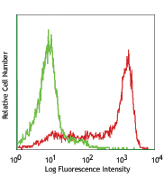
GFP-transfected CHO cells were fixed, permeabilized, and intracellularly stained with anti-GFP (clone FM264G) Purified (filled histogram) or rat IgG2a, κ isotype control (open histogram) followed by anti-rat IgG PE.
| Cat # | Size | Price | Save |
|---|---|---|---|
| 338001 | 25 µg | ¥15,620 | |
| 338002 | 100 µg | ¥28,380 |
Green fluorescent protein (GFP) was originally identified as a protein involved in bioluminescence, which is from the jellyfish Aequorea Victoria. It is widely used as a fluorescent indicator for monitoring gene expression in a variety of cellular systems, including living organisms and fixed tissues. Unlike other bioluminescent reporters, GFP fluoresces without the need for exogenous substrates or cofactors, or other intrinsic or extrinsic proteins, making GFP a useful tool for monitoring gene expression and protein localization in vivo. Purified GFP is a 27 kD monomer consisting of 238 amino acids and emits green light (emission maximum at 509 nm) when excited with blue or UV light.
Product DetailsProduct Details
- Antibody Type
- Monoclonal
- Host Species
- Rat
- Immunogen
- TLR9-GFP transfected cell line
- Formulation
- Phosphate-buffered solution, pH 7.2, containing 0.09% sodium azide.
- Preparation
- The antibody was purified by affinity chromatography.
- Concentration
- 0.5 mg/ml
- Storage & Handling
- The antibody solution should be stored undiluted between 2°C and 8°C.
- Application
-
ICFC - Quality tested
ICC - Reported in the literature, not verified in house - Recommended Usage
-
Each lot of this antibody is quality control tested by intracellular immunofluorescent staining with flow cytometric analysis. For flow cytometric staining, the suggested use of this reagent is ≤ 0.25 µg per 106 cells in 100 µl volume or 100 µl of whole blood. It is recommended that the reagent be titrated for optimal performance for each application.
- Application Notes
-
Additional reported applications (for the relevant formats) include: immunocytochemistry1.
-
Application References
(PubMed link indicates BioLegend citation) -
- Stephen LA, et al. 2018. Dev. Cell. 47(1):122-132.e4. PubMed (ICC)
- Product Citations
-
- RRID
-
AB_1279415 (BioLegend Cat. No. 338001)
AB_1279414 (BioLegend Cat. No. 338002)
Antigen Details
- Biology Area
- Cell Biology, Immunology
- Antigen References
-
1. Ishikura H, et al. 2004. Anticancer Res. 24:719.
2. Rizzuto R, et al. 1996. Curr. Biol. 6:183.
3. Chalfie M, et al. 1994. Science 263:802. - Gene ID
- NA
- UniProt
- View information about GFP on UniProt.org

Related FAQs
- Can I use common compensation control for GFP, CFSE and FITC because they emit in the same channel?
- It is not recommended even if they emit in the same channel because these are still different fluors with different brightness intensities. Individual compensation controls should be employed.
Other Formats
View All GFP Reagents Request Custom Conjugation| Description | Clone | Applications |
|---|---|---|
| Alexa Fluor® 488 anti-GFP | FM264G | ICFC,ICC |
| Purified anti-GFP | FM264G | ICFC,ICC |
| PE anti-GFP | FM264G | ICFC |
| Alexa Fluor® 647 anti-GFP | FM264G | ICFC,ICC |
| APC anti-GFP | FM264G | ICFC |
| PerCP/Cyanine5.5 anti-GFP | FM264G | ICFC |
| PE/Cyanine7 anti-GFP | FM264G | ICFC |
Customers Also Purchased

Compare Data Across All Formats
This data display is provided for general comparisons between formats.
Your actual data may vary due to variations in samples, target cells, instruments and their settings, staining conditions, and other factors.
If you need assistance with selecting the best format contact our expert technical support team.
-
Alexa Fluor® 488 anti-GFP

GFP transfected CHO cells were fixed and permeabilized, and ... -
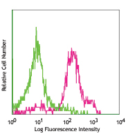
Purified anti-GFP

GFP-transfected CHO cells were fixed, permeabilized, and int... -
PE anti-GFP

GFP-transfected CHO cells were fixed and permeabilized, and ... -
Alexa Fluor® 647 anti-GFP

GFP transfected CHO cells intracellularly stained with FM264... -
APC anti-GFP

GFP-transfected CHO cells were fixed and permeabilized, and ... -
PerCP/Cyanine5.5 anti-GFP

GFP-transfected CHO cells were fixed and permeabilized, and ... -
PE/Cyanine7 anti-GFP

GFP-transfected RBL-1 cells were fixed and permeabilized, an...


Follow Us