- Clone
- 16E12C58 (See other available formats)
- Regulatory Status
- RUO
- Other Names
- CTSD, CPSD, CLN10, HEL-S-130P
- Isotype
- Mouse IgG1, κ
- Ave. Rating
- Submit a Review
- Product Citations
- publications

-
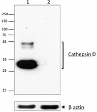
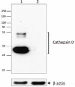
Total cell lysate from human MCF-7 (lane 1) and Raw264.7 cells (lane 2) were resolved by electrophoresis, transferred to nitrocellulose, and probed with purified monoclonal anti-Cathepsin D (clone 16E12C58) antibody. Proteins were visualized using an HRP-conjugated anti-mouse-IgG secondary and chemiluminescence detection.
Cathepsin D (CTSD) is a major lysosomal aspartic endoproteinase. It is active in acidic conditions with a pH range from 3.5 to 5.0, and its activity is inhibited by pepstatin A. CTSD is involved in lysosomal protein turnover and many other cellular processes. CTSD participates in the resolution of inflammation by initiating the apoptotic program in neutrophils. CTSD is released from neutrophil azurophilic granules during the initial stage of apoptosis, which leads to death receptor-independent activation of caspase-8. CTSD induces apoptosis in other cells as well. In T cells, CTSD activates Bax, which leads to the release of mitochondrial apoptosis-inducing factor (AIF), and subsequently induces an early caspase-independent apoptotic phenotype. CTSD also takes part in the release of cytochrome C from the mitochondria and activation of caspases in fibroblast apoptosis, which is induced by staurosporine. In addition, CTSD participates in cell proliferation, and it also increases proliferation and metastatic potential of many cancer cell lines. Overexpression of CTSD has been reported in many different cancers including malignant glioma, melanoma, breast, ovarian, endometrial, and bladder cancer. CTSD is also involved in neurodegenerative diseases. CTSD knockout mice die shortly after birth and display some neurodegeneration phenotypes. Mutations of CTSD cause congenital neuronal ceroid lipofuscinosis in humans and animals. CTSD cleaves the amyloid precursor protein and may play a role in Alzheimer’s disease. CTSD-deficiency has also been associated with Parkinson disease. Like CTSB and CTSL, CTSD functions outside the cells and is able to cleave extracellular matrix proteins including collagen, fibronectin, proteoglycans, and laminin.
Product DetailsProduct Details
- Verified Reactivity
- Human
- Antibody Type
- Monoclonal
- Host Species
- Mouse
- Immunogen
- Partial human cathepsin D recombinant protein (21-412 a.a.) expressed in 293E cells.
- Formulation
- Phosphate-buffered solution, pH 7.2, containing 0.09% sodium azide.
- Preparation
- The antibody was purified by affinity chromatography.
- Concentration
- 0.5 mg/ml
- Storage & Handling
- The antibody solution should be stored undiluted between 2°C and 8°C.
- Application
-
WB - Quality tested
- Recommended Usage
-
Each lot of this antibody is quality control tested by Western blotting. For Western blotting, the suggested use of this reagent is 0.2 - 1.0 µg per ml. It is recommended that the reagent be titrated for optimal performance for each application.
- RRID
-
AB_2565905 (BioLegend Cat. No. 678502)
Antigen Details
- Structure
- The 402 amino acid recombinant protein has a predicted molecular mass of approximately 44 kD. Aspartic endoproteinase.
- Distribution
- Lysosome. Secreted. CTSD is found in most cells and tissues of mammals. It is very abundant in macrophages.
- Function
- Lysosomal protein turnover, apoptosis, cell proliferation, cell invasion, neurodegeneration, and immune responses. Upregulated by estradiol, insulin, insulin-like growth factor, EGF, and FGF basic.
- Ligand/Receptor
- Caspase-8, collagen, fibronectin, proteoglycans, and laminin.
- Biology Area
- Apoptosis/Tumor Suppressors/Cell Death, Cell Biology, Neurodegeneration, Neuroscience, Neuroscience Cell Markers, Protein Trafficking and Clearance
- Molecular Family
- Enzymes and Regulators, Lysosomal Markers
- Antigen References
-
1. Vetvicka V, et al. 1994. Cancer Lett. 79:131.
2. Johansson AC, et al. 2003. Cell Death Differ. 10:1253.
3. Cavailles V, et al. 1988. Nucleic Acids Res. 16:1903.
4. Bidere N, et al. 2003. J. Biol. Chem. 278:31401.
5. Conus S, et al. 2012. J. Biol. Chem. 25:21142.
6. Koike M, et al. 2001. J. Neurosci. 21:7526.
7. Cullen V, et al. 2009. Mol. Brain 2:5.
8. Dian D, et al. 2014. Histol. Histopathol. 29:433. - Gene ID
- 1509 View all products for this Gene ID
- UniProt
- View information about Cathepsin D on UniProt.org
Related Pages & Pathways
Pages
Other Formats
View All Cathepsin D Reagents Request Custom Conjugation| Description | Clone | Applications |
|---|---|---|
| Purified anti-Cathepsin D | 16E12C58 | WB |
Compare Data Across All Formats
This data display is provided for general comparisons between formats.
Your actual data may vary due to variations in samples, target cells, instruments and their settings, staining conditions, and other factors.
If you need assistance with selecting the best format contact our expert technical support team.
-
Purified anti-Cathepsin D
Total cell lysate from human MCF-7 (lane 1) and Raw264.7 cel...


Follow Us