


- Clone
- DECMA-1 (See other available formats)
- Regulatory Status
- RUO
- Other Names
- E-Cadherin, Cadherin-1, CDH1, and UVO
- Isotype
- Rat IgG1, κ
- Ave. Rating
- Submit a Review
- Product Citations
- publications

-

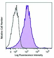
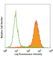
MDCK epithelial cell line was stained with CD324 (clone DECMA-1) PE/Cyanine7 (filled histogram) or rat IgG1, ? PE/Cyanine7 isotype control (open histogram).
| Cat # | Size | Price | Save |
|---|---|---|---|
| 147309 | 25 µg | ¥27,280 | |
| 147310 | 100 µg | ¥81,400 |
CD324, also known as E-cadherin, cadherin-1, CDH1, and UVO is a member of the cadherin superfamily. It is a calcium-dependent, transmembrane cell-cell adhesion glycoprotein composed of four extracellular cadherin repeats and a highly conserved cytoplasmic tail region. CD324 is widely expressed in epithelial cells in the colon, uterus, liver, keratinocytes, brain, heart, muscle, kidney, and pancreas as well as erythroid cells. CD324 functions as a cell adhesion molecule involved in development, bacterial pathogenesis, and tumor invasion. In bacterial pathogenesis, the ectodomain of CD324 mediates bacterial adhesion to mammalian cells, while the cytoplasmic domain is required for internalization. CD324 binds to the αEβ7 integrin to mediate cell adhesion and also interacts with a number of intracellular proteins including including erbin, ezrin, caspase-3, caspase-8, β-catenin, presenilin 1, and casein kinase II as well as other extracellular proteins including the EGF receptor.
Product DetailsProduct Details
- Verified Reactivity
- Mouse, Human
- Reported Reactivity
- Cynomolgus, Dog, Pig
- Antibody Type
- Monoclonal
- Host Species
- Rat
- Immunogen
- E-Cadherin extracellular domain
- Formulation
- Phosphate-buffered solution, pH 7.2, containing 0.09% sodium azide.
- Preparation
- The antibody was purified by affinity chromatography and conjugated with PE/Cyanine7 under optimal conditions.
- Concentration
- 0.2 mg/ml
- Storage & Handling
- The antibody solution should be stored undiluted between 2°C and 8°C, and protected from prolonged exposure to light. Do not freeze.
- Application
-
FC - Quality tested
- Recommended Usage
-
Each lot of this antibody is quality control tested by immunofluorescent staining with flow cytometric analysis. For flow cytometric staining, the suggested use of this reagent is =0.25 µg per million cells in 100 µl volume. It is recommended that the reagent be titrated for optimal performance for each application.
It is also recommended using EDTA-based solutions for dissociating attachment-dependent cell lines. - Excitation Laser
-
Blue Laser (488 nm)
Green Laser (532 nm)/Yellow-Green Laser (561 nm)
- Application Notes
-
Additional reported applications (for relevant formats) include: immunoprecipitation1, Western Blotting1, immunomicroscopy3, biological function1,2, and spatial biology (IBEX)4,5.
- Additional Product Notes
-
BioLegend is in the process of converting the name PE/Cy7 to PE/Cyanine7. The dye molecule remains the same, so you should expect the same quality and performance from our PE/Cyanine7 products. Please contact Technical Service if you have any questions.
-
Application References
(PubMed link indicates BioLegend citation) -
- Vestweber D, et al. 1985. EMBO. 4:3393. (IP, WB, FA)
- Nakagawa M, et al. 2001. J. Cell Sci. 114:1829. (FA in canine cells)
- Mohamet L, et al. 2010. PLoS ONE. 5:e12921. (IF)
- Radtke AJ, et al. 2020. Proc Natl Acad Sci U S A. 117:33455-65. (SB) PubMed
- Radtke AJ, et al. 2022. Nat Protoc. 17:378-401. (SB) PubMed
- Product Citations
-
- RRID
-
AB_2564187 (BioLegend Cat. No. 147309)
AB_2564188 (BioLegend Cat. No. 147310)
Antigen Details
- Structure
- Member of the cadherin superfamily. Calcium-dependent, transmembrane cell-cell adhesion glycoprotein composed of four extracellular cadherin repeats and a highly conserved cytoplasmic tail region.
- Distribution
-
Widely expressed in epithelial cells in the colon, uterus, liver, keratinocytes, brain, heart, muscle, kidney, and pancreas as well as erythroid cells.
- Function
- Cell adhesion molecule involved in development, bacterial pathogenesis, and tumor invasion. The ectodomain of CD324 mediates bacterial adhesion to mammalian cells, while the cytoplasmic domain is required for internalization.
- Interaction
- Interacts with a variety of proteins including erbin, ezrin, caspase-3, caspase-8, EGF receptor, β-catenin, presenilin 1, casein kinase II, and others.
- Ligand/Receptor
- αEβ7 integrin.
- Cell Type
- Embryonic Stem Cells
- Biology Area
- Cell Adhesion, Cell Biology, Immunology, Innate Immunity, Neuroscience, Stem Cells, Synaptic Biology
- Molecular Family
- Adhesion Molecules, CD Molecules
- Antigen References
-
1. Overduin M, et al. 1995. Science 267:386.
2. Boggon TJ, et al. 2002. Science 296:1308.
3. Berx G, et al. 1995. EMBO J. 14:6107.
4. Perl AK, et al. 1998. Nature 392:190. - Gene ID
- 999 View all products for this Gene ID 12550 View all products for this Gene ID
- UniProt
- View information about CD324 on UniProt.org
Related Pages & Pathways
Pathways


Related FAQs
Other Formats
View All CD324 Reagents Request Custom Conjugation| Description | Clone | Applications |
|---|---|---|
| Purified anti-mouse/human CD324 (E-Cadherin) | DECMA-1 | FC,ICC,IP,WB,FA |
| PE anti-mouse/human CD324 (E-Cadherin) | DECMA-1 | FC |
| Alexa Fluor® 594 anti-mouse/human CD324 (E-Cadherin) | DECMA-1 | ICC,IHC-P,IHC-F,3D IHC |
| Alexa Fluor® 647 anti-mouse/human CD324 (E-Cadherin) | DECMA-1 | FC,ICC,IHC-F,3D IHC,SB |
| PE/Cyanine7 anti-mouse/human CD324 (E-Cadherin) | DECMA-1 | FC |
| PE/Dazzle™ 594 anti-mouse/human CD324 (E-Cadherin) | DECMA-1 | FC |
| PerCP/Cyanine5.5 anti-mouse/human CD324 (E-Cadherin) | DECMA-1 | FC |
| APC anti-mouse/human CD324 (E-Cadherin) | DECMA-1 | FC |
| Brilliant Violet 421™ anti-mouse/human CD324 (E-Cadherin) | DECMA-1 | FC |
| APC/Fire™ 750 anti-mouse/human CD324 (E-Cadherin) | DECMA-1 | FC |
Customers Also Purchased
Compare Data Across All Formats
This data display is provided for general comparisons between formats.
Your actual data may vary due to variations in samples, target cells, instruments and their settings, staining conditions, and other factors.
If you need assistance with selecting the best format contact our expert technical support team.
-
Purified anti-mouse/human CD324 (E-Cadherin)

Canine kidney cell line MDCK was cultured in a chamber slide... 
MDCK epithelial cell line was stained with purified CD324 (c... -
PE anti-mouse/human CD324 (E-Cadherin)

MDCK epithelial cell line was stained with CD324 (clone DECM... -
Alexa Fluor® 594 anti-mouse/human CD324 (E-Cadherin)

Madin-darby canine kidney epithelial cell line, MDCK, was cu... 
C57BL/6 mouse frozen intestine section was fixed with 4% par... 
Human paraffin-embedded colon cancer tissue slices were prep... 
Paraformaldehyde-fixed (1%), 500 μm-thick mouse liver tissue... -
Alexa Fluor® 647 anti-mouse/human CD324 (E-Cadherin)

MDCK epithelial cell line was stained with CD324 (clone DECM... 
Madin-darby canine kidney epithelial cell line, MDCK, was cu... 
C57BL/6 mouse frozen intestine section was fixed with 4% par... 
Paraformaldehyde-fixed (4%), 500 μm-thick mouse lung tissue ... 
Confocal image of C57BL/6 mouse liver sample acquired using ... 
Confocal image of C57BL/6 mouse liver sample acquired using ... -
PE/Cyanine7 anti-mouse/human CD324 (E-Cadherin)

MDCK epithelial cell line was stained with CD324 (clone DECM... -
PE/Dazzle™ 594 anti-mouse/human CD324 (E-Cadherin)

MDCK epithelial cell line was stained with CD324 (E-Cadherin... -
PerCP/Cyanine5.5 anti-mouse/human CD324 (E-Cadherin)

MDCK epithelial cell line was stained with CD324 (E-Cadherin... -
APC anti-mouse/human CD324 (E-Cadherin)

MDCK epithelial cell line was stained with CD324 (clone DECM... -
Brilliant Violet 421™ anti-mouse/human CD324 (E-Cadherin)

MDCK epithelial cell line was stained with CD324 (E-Cadherin... -
APC/Fire™ 750 anti-mouse/human CD324 (E-Cadherin)

MDCK epithelial cell line was stained with CD324 (clone DECM...


Follow Us