


- Clone
- 9C10 (MFR4.B) (See other available formats)
- Regulatory Status
- RUO
- Other Names
- α4 integrin, VLA-4 α chain, integrin α4, ITGA4
- Isotype
- Rat IgG2a, κ
- Ave. Rating
- Submit a Review
- Product Citations
- publications

-

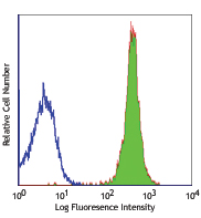
C57BL/6 mouse splenocytes stained with 9C10 PE
| Cat # | Size | Price | Save |
|---|---|---|---|
| 103705 | 50 µg | ¥28,380 | |
| 103706 | 200 µg | ¥66,880 |
CD49d is a 150 kD glycoprotein, also known as α4 integrin or VLA-4 α chain. It is a member of the integrin family, expressed on T and B cells, monocytes, eosinophils, basophils, mast cells, thymocytes, NK cells, and dendritic cells. CD49d is a heterodimer expressed with either of two β chains, β1 (CD29) or β7, to form the VLA-4 (integrin α4β1) or LPAM-1 (integrin α4β7) complexes. CD49d plays a critical role in both adhesion and T cell costimulation. The primary ligands for CD49d are VCAM-1, MAdCAM-1, and fibronectin. The 9C10(MFR4.B) antibody, in combination with the R1-2 monoclonal antibody, completely blocks VCAM-1 binding to VLA-4.
Product DetailsProduct Details
- Verified Reactivity
- Mouse
- Antibody Type
- Monoclonal
- Host Species
- Rat
- Immunogen
- (C57BL/6 x A/J)F1 mouse fetal liver mas
- Formulation
- Phosphate-buffered solution, pH 7.2, containing 0.09% sodium azide.
- Preparation
- The antibody was purified by affinity chromatography, and conjugated with PE under optimal conditions.
- Concentration
- 0.2 mg/ml
- Storage & Handling
- The antibody solution should be stored undiluted between 2°C and 8°C, and protected from prolonged exposure to light. Do not freeze.
- Application
-
FC - Quality tested
- Recommended Usage
-
Each lot of this antibody is quality control tested by immunofluorescent staining with flow cytometric analysis. For flow cytometric staining, the suggested use of this reagent is ≤ 0.25 µg per 106 cells in 100 µl volume. It is recommended that the reagent be titrated for optimal performance for each application.
- Excitation Laser
-
Blue Laser (488 nm)
Green Laser (532 nm)/Yellow-Green Laser (561 nm)
- Application Notes
-
Additional reported applications (for the relevant formats) include: in vitro blocking of cell-cell adhesion and T cell costimulation1,2, and immunohistochemistry3 of acetone-fixed frozen sections. The Ultra-LEAF™ purified antibody (Endotoxin <0.01 EU/µg, Azide-Free, 0.2 µm filtered) is recommended for functional assays (Cat. No. 103709 & 103710).
-
Application References
(PubMed link indicates BioLegend citation) -
- Kinashi T, et al. 1994. Blood Cells 20:25. (Block)
- Halvorson MJ, et al. 1995. J. Immunol. 155:4567. (Block)
- Hata H, et al. 2004. J. Clin. Invest. 114:582. (IHC)
- Jia W, et al. 2005. Blood 106:3854. (FC)
- Product Citations
-
- RRID
-
AB_313046 (BioLegend Cat. No. 103705)
AB_313047 (BioLegend Cat. No. 103706)
Antigen Details
- Structure
- Integrin family, associates with integrin β1 or β7, 150 kD
- Distribution
-
T cells and B cells, monocytes, eosinophils, basophils, mast cells, thymocytes, NK cells, dendritic cells
- Function
- T cell costimulation, cell-cell and cell-matrix adhesion
- Ligand/Receptor
- VCAM-1, MAdCAM-1, fibronectin
- Cell Type
- B cells, Basophils, Dendritic cells, Eosinophils, Mast cells, Monocytes, NK cells, T cells, Thymocytes, Tregs
- Biology Area
- Cell Adhesion, Cell Biology, Immunology, Innate Immunity
- Molecular Family
- Adhesion Molecules, CD Molecules
- Antigen References
-
1. Barclay AN, et al. 1997. The Leukocyte Antigen FactsBook Academic Press.
2. Lobb RR, et al. 1994. J. Clin. Invest. 94:1722.
3. Berlin C, et al. 1993. Cell 74:185.
4. Maguire JE, et al. 1995. J. Exp. Med. 182:2079. - Gene ID
- 16401 View all products for this Gene ID
- UniProt
- View information about CD49d on UniProt.org
Related Pages & Pathways
Pathways


Related FAQs
- What type of PE do you use in your conjugates?
- We use R-PE in our conjugates.
Other Formats
View All CD49d Reagents Request Custom Conjugation| Description | Clone | Applications |
|---|---|---|
| Biotin anti-mouse CD49d | 9C10 (MFR4.B) | FC |
| PE anti-mouse CD49d | 9C10 (MFR4.B) | FC |
| Purified anti-mouse CD49d | 9C10 (MFR4.B) | FC,Block,IHC-F |
| Ultra-LEAF™ Purified anti-mouse CD49d | 9C10 (MFR4.B) | FC,Block,IHC-F |
Customers Also Purchased
Compare Data Across All Formats
This data display is provided for general comparisons between formats.
Your actual data may vary due to variations in samples, target cells, instruments and their settings, staining conditions, and other factors.
If you need assistance with selecting the best format contact our expert technical support team.
-
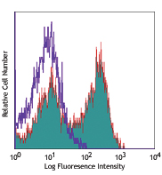
Biotin anti-mouse CD49d

C57BL/6 mouse splenocytes stained with 9C10 biotin and detec... -
PE anti-mouse CD49d

C57BL/6 mouse splenocytes stained with 9C10 PE -
Purified anti-mouse CD49d

C57BL/6 mouse splenocytes stained with purified 9C10, follow... -
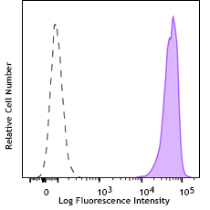
Ultra-LEAF™ Purified anti-mouse CD49d

C57BL/6 splenocytes stained with Ultra-LEAF™ purified 9C10, ...


Follow Us