


- Clone
- BL168; UCHT1; RPA-T4;
- Regulatory Status
- RUO
- Other Names
- Th17 kit, Th-17 kit, Th 17 kit
- Isotype
- Mouse IgG1, κ (all clones)
- Ave. Rating
- Submit a Review
- Product Citations
- publications

-

CD3 FITC -

-

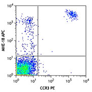
PMA/ionomycin-stimulated (6hr) human peripheral blood lymphocytes stained with Human Th17 flow(TM) kit. Dot plot analysis is derived from gated in CD3+ cell population.
Th17 cells are a subset of CD4+ T cells defined by their ability to produce the cytokines IL-17, IL-17F and IL-22. Th17 cells participate in normal epithelial immunity, their dysregulation is implicated in psoriasis, multiple sclerosis, systemic lupus erythmatosis, rheumatoid arthritis, and inflammatory bowel disease. BioLegend’s human Th17 flow kit is designed and formulated specifically for one-step intracellular immunofluorescence staining and flow cytometric analysis of Th17 cells in a mixed cell population. This kit is composed of fluorochrome conjugated anti-human CD3, CD4 and IL-17 antibodies, isotype control and the critical buffers. It is easy to use for identification of Th17 cells.
Materials provided:
- Mouse IgG1,k isotype control Alexa Fluor® 647/CD3 FITC/CD4 PE,25 tests
- Anti-human IL-17 Alexa Fuor® 647/CD3 FITC/CD4 PE, 25 tests
- Fixation Buffer, 100ml, Cat. No. 420801
- Intracellular Staining Perm Wash Buffer (10X), 100 ml, Cat. No. 421002
Materials not included:
- Cell Staining Buffer (Cat. No. 420201)
Product Details
- Verified Reactivity
- Human
- Host Species
- Mouse
- Concentration
- Lot-specific (to obtain lot-specific concentration and expiration, please enter the lot number in our Certificate of Analysis online tool.)
- Storage & Handling
-
Upon receipt, store between 2°C and 8°C, and protected from prolonged exposure to light. Do not freeze. To obtain lot-specific expiration date, please enter the lot number in our Concentration and Expiration Lookup or Certificate of Analysis online tools.)
This product has a shelf-life of 12 months or less. Please contact our technical support team for lot specific CoA and expiration date inquiries of this product. - Application
-
ICFC - Quality tested
- Application Notes
-
Staining Procedure:
1. Distribute 0.5-1 X 10⁶ cells/100 µl/tube into 12 X 75 mm plastic testing tubes.
2. Add 0.5 ml/tube Fixation Buffer (Cat. No. 420801) to each tube, votex and incubate at room temperature for 20 minutes.
3. Centrifuge at 350 x g for 5 minutes, discard supernatant.
4. Prepare 1X Intracellular Staining Perm Wash Buffer (Cat. No. 421002) by diluting 1 part of 10X Intracellular Staining Perm Wash Buffer (Cat. No. 421002) with 9 part of DI water.
5. Add 2ml 1X Intracellular Staining Perm Wash Buffer to each tube, vortex and incubate at room temperature for 20 minutes, then spin at 350 x g for 5 minutes and remove the supernatant.
6. Wash once by adding 2ml 1X Intracellular Staining Perm Wash Buffer to each tube, spin and remove the supernatant.
7. Resuspend the pellet in 100 µl of 1X Intracellular Staining Perm Wash Buffer.
8. Add 20 µl Alexa Fluor® 647 mouse IgG1, k isotype control/CD3 FITC/CD4 PE or IL-17 Alexa Fluor® 647/CD3 FITC/CD4 PE into appropriate tube and incubate at room temperature in the dark for 30 minutes.
10. Wash twice with 2ml 1X Intracellular Staining Perm Wash Buffer, and resuspend in 0.5ml cell staining buffer then analyze under flow cytometer with appropriate instrument setting.
Note:
1. The Fixation buffer contains 4% paraformaldehyde, which is toxic and is a suspected carcinogen. Contact with eyes, skin and mucous membranes should be avoided.
2. Alexa Fluor® is a registered trademark of Molecular Probes, Inc. Alexa Fluor® dye antibody conjugates are sold under license from Molecular Probes, Inc. for research use only, except for use in combination with microarrays and high content screening, and are covered by pending and issued patents.
3. Please note that single color controls are required to perform instrument compensation and are not included in the kit. -
Application References
(PubMed link indicates BioLegend citation) - Product Citations
-
Antigen Details
- Biology Area
- Immunology
- Molecular Family
- Cytokines/Chemokines
- Antigen References
-
1. King C, et al. 2004 Cell 117:265.
2. Betelli E, et al. 2006 Nature 441:235.
3. Betelli E, et al. 2007 Nat. Imm. 8:345.
4. Wilson NJ, et al. 2007 Nat. Imm. 8:950. - Gene ID
- 3605 View all products for this Gene ID
- UniProt
- View information about Th17 on UniProt.org
Related Pages & Pathways
Pathways


Related FAQs
Other Formats
View All Reagents Request Custom Conjugation| Description | Clone | Applications |
|---|---|---|
| Human Th17 Flow™ Kit (CD3 FITC/CD4 PE/IL-17 Alexa Fluor® 647) | BL168; UCHT1; RPA-T4 | ICFC |
Customers Also Purchased
_121609.jpg)
Compare Data Across All Formats
This data display is provided for general comparisons between formats.
Your actual data may vary due to variations in samples, target cells, instruments and their settings, staining conditions, and other factors.
If you need assistance with selecting the best format contact our expert technical support team.
-
Human Th17 Flow™ Kit (CD3 FITC/CD4 PE/IL-17 Alexa Fluor® 647)

CD3 FITC 
PMA/ionomycin-stimulated (6hr) human peripheral blood lympho... 


Follow Us