


- Clone
- RM4-5 (See other available formats)
- Regulatory Status
- RUO
- Other Names
- L3T4, T4
- Isotype
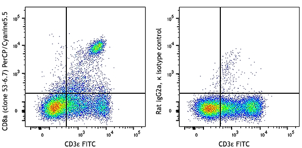
- Rat IgG2a, κ
- Ave. Rating
- Submit a Review
- Product Citations
- publications

-

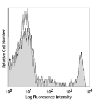
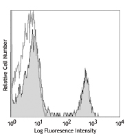
C57BL/6 mouse splenocytes stained with CD4 (clone RM4-5) FITC (solid line) or rat IgG2a, κ FITC isotype control (dotted line).
| Cat # | Size | Price | Save |
|---|---|---|---|
| 100509 | 50 µg | ¥6,600 | |
| 100510 | 500 µg | ¥20,700 |
CD4 is a 55 kD protein also known as L3T4 or T4. It is a member of the Ig superfamily, primarily expressed on most thymocytes and a subset of T cells, and weakly on macrophages and dendritic cells. It acts as a co-receptor with the TCR during T cell activation and thymic differentiation by binding MHC class II and associating with the protein tyrosine kinase lck.
Product DetailsProduct Details
- Verified Reactivity
- Mouse
- Antibody Type
- Monoclonal
- Host Species
- Rat
- Immunogen
- BALB/c mouse thymocytes
- Formulation
- Phosphate-buffered solution, pH 7.2, containing 0.09% sodium azide.
- Preparation
- The antibody was purified by affinity chromatography, and conjugated with FITC under optimal conditions.
- Concentration
- 0.5 mg/ml
- Storage & Handling
- The antibody solution should be stored undiluted between 2°C and 8°C, and protected from prolonged exposure to light. Do not freeze.
- Application
-
FC - Quality tested
- Recommended Usage
-
Each lot of this antibody is quality control tested by immunofluorescent staining with flow cytometric analysis. For flow cytometric staining, the suggested use of this reagent is ≤0.25 µg per million cells in 100 µl volume. It is recommended that the reagent be titrated for optimal performance for each application.
- Excitation Laser
-
Blue Laser (488 nm)
- Application Notes
-
The RM4-5 antibody blocks the binding of GK1.5 antibody and H129.19 antibody to CD4+ T cells, but not RM4-4 antibody. Additional reported applications (for the relevant formats) include: blocking of ligand binding, in vivo depletion of CD4+ cells1, and immunohistochemistry of acetone-fixed frozen tissue sections2,3,11 and paraffin-embedded sections11. Clone RM4-5 is not recommended for immunohistochemistry of formalin-fixed paraffin sections. Instead, acetone frozen or zinc-fixed paraffin sections are recommended. The Ultra-LEAF™ Purified antibody (Endotoxin < 0.01 EU/µg, Azide-Free, 0.2 µm filtered) is recommended for functional assays (Cat. No. 100575 and 100576).
-
Application References
(PubMed link indicates BioLegend citation) -
- Kruisbeek AM. 1991. In Curr. Protocols Immunol. pp. 4.1.1-4.1.5. (Block, Deplete)
- Nitta H, et al. 1997. Cell Vision 4:73. (IHC)
- Fan WY, et al. 2001. Exp. Biol. Med. 226:1045.
- Muraille E, et al. 2003. Infect. Immun. 71:2704. (IHC)
- León-Ponte M, et al. 2007. Blood 109:3139. (FC)
- Bourdeau A, et al. 2007. Blood doi:10.1182/blood-2006-08-044370. (FC)
- Matsumoto M, et al. 2007.J. Immunol.178:2499. PubMed
- Shigeta A, et al. 2008. Blood 112:4915. PubMed
- Zaborsky N, et al. 2010. J. Immunol. 184:725. PubMed
- Rodrigues-Manzanet R, et al. 2010. P. Natl Acad Sci USA 107:8706. PubMed
- Whiteland JL, et al. 1995. J. Histochem. Cytochem. 43:313. (IHC)
- Product Citations
-
- RRID
-
AB_312712 (BioLegend Cat. No. 100509)
AB_312713 (BioLegend Cat. No. 100510)
Antigen Details
- Structure
- Ig superfamily, 55 kD
- Distribution
-
Majority of thymocytes, T cell subset
- Function
- TCR co-receptor, T cell activation
- Ligand/Receptor
- MHC class II molecule
- Cell Type
- Dendritic cells, T cells, Thymocytes, Tregs
- Biology Area
- Immunology
- Molecular Family
- CD Molecules
- Antigen References
-
1. Barclay A, et al. 1997. The Leukocyte Antigen FactsBook Academic Press.
2. Bierer BE, et al. 1989. Annu. Rev. Immunol. 7:579.
3. Janeway CA. 1992. Annu. Rev. Immunol. 10:645. - Gene ID
- 12504 View all products for this Gene ID
- UniProt
- View information about CD4 on UniProt.org
Related Pages & Pathways
Pathways


Related FAQs
- I am unable to see expression of T cell markers such as CD3 and CD4 post activation.
- TCR-CD3 complexes on the T-lymphocyte surface are rapidly downregulated upon activation with peptide-MHC complex, superantigen or cross-linking with anti-TCR or anti-CD3 antibodies. PMA/Ionomycin treatment has been shown to downregulate surface CD4 expression. Receptor downregulation is a common biological phenomenon and so make sure that your stimulation treatment is not causing it in your sample type.
Other Formats
View All CD4 Reagents Request Custom ConjugationCustomers Also Purchased
Compare Data Across All Formats
This data display is provided for general comparisons between formats.
Your actual data may vary due to variations in samples, target cells, instruments and their settings, staining conditions, and other factors.
If you need assistance with selecting the best format contact our expert technical support team.
-
APC anti-mouse CD4

C57BL/6 mouse splenocytes stained with CD4 (clone RM4-5) APC... -
Biotin anti-mouse CD4

C57BL/6 mouse splenocytes stained with biotinylated CD4 (clo... -
FITC anti-mouse CD4

C57BL/6 mouse splenocytes stained with CD4 (clone RM4-5) FIT... -
PE anti-mouse CD4

C57BL/6 mouse splenocytes stained with CD4 (clone RM4-5) PE ... 
C57BL/6 mouse splenocytes were stained with CD4 (clone RM4-5... -
PE/Cyanine5 anti-mouse CD4

C57BL/6 mouse splenocytes stained with CD4 (clone RM4-5) PE/... -
Purified anti-mouse CD4

C57BL/6 mouse splenocytes stained with purified CD4 (clone R... 
C57BL/6 mouse frozen spleen section was fixed with 4% parafo... -
PE/Cyanine7 anti-mouse CD4

C57BL/6 mouse splenocytes stained with CD4 (clone RM4-5) PE/... -
APC/Cyanine7 anti-mouse CD4

C57BL/6 mouse splenocytes stained with CD4 (clone RM4-5) APC... -
Alexa Fluor® 647 anti-mouse CD4

C57BL/6 mouse splenocytes stained with CD4 (clone RM4-5) Ale... Formalin-fixed, 300 micron-thick mouse spleen section was bl... 
C57BL/6 mouse frozen spleen section was fixed with 4% parafo... 
Paraformaldehyde-fixed (4%), 500 μm-thick mouse spleen tissu... -
Alexa Fluor® 488 anti-mouse CD4

C57BL/6 mouse splenocytes stained with CD4 (clone RM4-5) Ale... 
C57BL/6 mouse frozen spleen section was fixed with 4% parafo... 
Paraformaldehyde-fixed (4%), 500 μm-thick mouse thymus tissu... -
Pacific Blue™ anti-mouse CD4

C57BL/6 mouse splenocytes stained with CD4 (clone RM4-5) Pac... -
Alexa Fluor® 700 anti-mouse CD4

C57BL/6 mouse splenocytes stained with CD4 (clone RM4-5) Ale... -
PerCP anti-mouse CD4

C57BL/6 mouse splenocytes stained with CD4 (clone RM4-5) Per... -
PerCP/Cyanine5.5 anti-mouse CD4

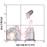
C57BL/6 mouse splenocytes stained with CD3e APC and CD4 (clo... -
Brilliant Violet 421™ anti-mouse CD4

C57BL/6 mouse splenocytes were stained with CD3 APC and CD4 ... 

C57BL/6 mouse thymocytes were fixed with 1% paraformaldehyde... 
C57BL/6 mouse frozen spleen section was fixed with 4% parafo... -
APC/Fire™ 750 anti-mouse CD4

C57BL/6 splenocytes were stained with CD3 FITC and CD4 (clon... 
-
Brilliant Violet 570™ anti-mouse CD4


-
Brilliant Violet 605™ anti-mouse CD4

C57BL/6 mouse splenocytes were stained with CD3 PE and CD4 (... -
Brilliant Violet 650™ anti-mouse CD4

C57BL/6 mouse splenocytes were stained with CD3 PE and CD4 (... -
Brilliant Violet 711™ anti-mouse CD4

C57BL/6 mouse splenocytes were stained with CD3 PE and CD4 (... -
Brilliant Violet 785™ anti-mouse CD4

C57BL/6 mouse splenocytes were stained with CD3 PE and CD4 (... -
Brilliant Violet 510™ anti-mouse CD4

C57BL/6 mouse splenocytes were stained with CD3 APC and CD4 ... -
Purified anti-mouse CD4 (Maxpar® Ready)

C57BL/6 mouse splenocytes stained with 147Sm-anti-CD45 (30-F... -
PE/Dazzle™ 594 anti-mouse CD4

C57BL/6 mouse splenocytes were stained with CD4 (clone RM4-5... -
TotalSeq™-A0001 anti-mouse CD4
-
TotalSeq™-B0001 anti-mouse CD4
-
TotalSeq™-C0001 anti-mouse CD4
-
Ultra-LEAF™ Purified anti-mouse CD4

C57BL/6 mouse splenocytes stained with LEAF™ purified CD4 (c... -
Spark Violet™ 423 anti-mouse CD4 (L3T4) Antibody

C57BL/6 splenocytes were stained with anti-mouse CD3e PE and... -
Spark Red™ 718 anti-mouse CD4 (L3T4) (Flexi-Fluor™)


Follow Us