


- Clone
- SK7 (See other available formats)
- Regulatory Status
- RUO
- Workshop
- HCDM listed
- Other Names
- T3, CD3ε
- Isotype
- Mouse IgG1, κ
- Ave. Rating
- Submit a Review
- Product Citations
- publications

-

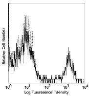
Human peripheral blood lymphocytes stained with biotinylated anti-human CD3 (clone SK7, filled histogram) or biotinylated mouse IgG1, κ (open histogram) isotype control, followed by SAV-PE -

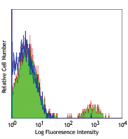
Human PBMCs, stimulated with 1 µg/ml of LPS for 8 h and treated with Brefeldin A during the last 4 h, were prepared by cytospin, fixed and permeabilized on a slide and then treated with endogenous biotin blocking kit (Vector labs). Slides were stained with anti-human CD3 biotin (clone SK7) and DyLight™ 649 streptavidin (green) and counterstained with DAPI (blue). Images were acquired with an Olympus FV10i confocal microscope. Images courtesy of Teresa Rodriguez, Darius Schneider, and Matthias von Herrath, LaJolla Institute for Allergy and Immunology.
CD3ε is a 20 kD chain of the CD3/T-cell receptor (TCR) complex, which is composed of two CD3ε, one CD3γ, one CD3δ, one CD3ζ (CD247), and a T-cell receptor (α/β or γ/δ) heterodimer. It is found on all mature T cells, NK T cells, and some thymocytes. CD3, also known as T3, is a member of the immunoglobulin superfamily that plays a role in antigen recognition, signal transduction, and T cell activation.
Product DetailsProduct Details
- Verified Reactivity
- Human
- Reported Reactivity
- Chimpanzee
- Antibody Type
- Monoclonal
- Host Species
- Mouse
- Formulation
- Phosphate-buffered solution, pH 7.2, containing 0.09% sodium azide.
- Preparation
- The antibody was purified by affinity chromatography, and conjugated with biotin under optimal conditions.
- Concentration
- 0.5 mg/ml
- Storage & Handling
- The antibody solution should be stored undiluted between 2°C and 8°C. Do not freeze.
- Application
-
FC - Quality tested
ICC - Verified - Recommended Usage
-
Each lot of this antibody is quality control tested by immunofluorescent staining with flow cytometric analysis. For flow cytometric staining, the suggested use of this reagent is ≤ 0.5 µg per million cells in 100 µl volume. It is recommended that the reagent be titrated for optimal performance for each application.
- Application Notes
-
Additional reported application (for the relevant formats) include: immunohistochemical staining of frozen tissue sections4,5,8, immunofluorescent staining6, and Western blotting3.
-
Application References
(PubMed link indicates BioLegend citation) -
- Kan EA, et al. 1983. J. Immunol. 131:536.
- Wood GS, et al. 1985. Am. J. Pathol. 120:371.
- Van Dongen JJM, et al. 1988. Blood 71:603. (WB)
- Haringman JJ, et al. 2005. Arthritis Res. Ther. 7:R862. (IHC)
- Carbone A, et al. 1999. Blood 93:2319. (IHC)
- Goval JJ, et al. 2006. J. Histochem. Cytochem. 54:75. (IF)
- Rutjens E, et al. 2007. J. Immunol. 178:1702.
- Kap Y, et al. 2009. J. Histochem. Cytochem. 57:1159. (IHC)
- Yoshino N, et al. 2000. Exp. Anim. (Tokyo) 49:97. (FC)
- Product Citations
-
- RRID
-
AB_10662538 (BioLegend Cat. No. 344820)
Antigen Details
- Structure
- Ig superfamily, with the subunits of CD3γ, CD3δ, CD3ζ, (CD247) and TCR (α/β or γ/δ) forms CD3/TCR complex, 20 kD
- Distribution
-
Mature T and NK T cells, during thymocyte differentiation
- Function
- Antigen recognition, signal transduction, T cell activation
- Ligand/Receptor
- Peptide antigen bound to MHC
- Cell Type
- NKT cells, T cells, Tregs
- Biology Area
- Immunology, Innate Immunity
- Molecular Family
- CD Molecules, TCRs
- Antigen References
-
1. Barclay N, et al. 1993. The Leucocyte FactsBook. Academic Press. San Diego.
2. Beverly P, et al. 1981. Eur. J. Immunol. 11:329.
3. Lanier L, et al. 1986. J. Immunol. 137:2501. - Gene ID
- 916 View all products for this Gene ID
- UniProt
- View information about CD3 on UniProt.org
Related Pages & Pathways
Pathways


Related FAQs
- How many biotin molecules are per antibody structure?
- We don't routinely measure the number of biotins with our antibody products but the number of biotin molecules range from 3-6 molecules per antibody.
Other Formats
View All CD3 Reagents Request Custom ConjugationCustomers Also Purchased

Compare Data Across All Formats
This data display is provided for general comparisons between formats.
Your actual data may vary due to variations in samples, target cells, instruments and their settings, staining conditions, and other factors.
If you need assistance with selecting the best format contact our expert technical support team.
-
APC/Fire™ 750 anti-human CD3

Human peripheral blood lymphocytes were stained with CD19 PE... 
-
Biotin anti-human CD3

Human peripheral blood lymphocytes stained with biotinylated... 
Human PBMCs, stimulated with 1 µg/ml of LPS for 8 h and trea... -
Purified anti-human CD3

Human peripheral blood lymphocytes stained with SK7 FITC. -
FITC anti-human CD3
Human peripheral blood lymphocytes stained with SK7 FITC. -
PE anti-human CD3

Human peripheral blood lymphocytes were stained with CD3 (cl... -
Alexa Fluor® 488 anti-human CD3

Human peripheral blood lymphocytes stained with SK7 Alexa Fl... -
APC anti-human CD3

Human peripheral blood lymphocytes stained with SK7 APC -
PerCP/Cyanine5.5 anti-human CD3

Human peripheral blood lymphocytes were stained with CD3 (cl... -
PerCP anti-human CD3

Human peripheral blood lymphocytes stained with SK7 PerCP -
PE/Cyanine7 anti-human CD3

Human peripheral blood lymphocytes stained with SK7 PE/Cyani... -
APC/Cyanine7 anti-human CD3

Human peripheral blood lymphocytes were stained with CD3 (cl... -
Alexa Fluor® 700 anti-human CD3

Human peripheral blood lymphocytes were stained with CD3 (cl... -
Pacific Blue™ anti-human CD3

Human peripheral blood lymphocytes were stained with CD3 (cl... -
Alexa Fluor® 647 anti-human CD3

Human peripheral blood lymphocytes were stained with CD3 (cl... -
Brilliant Violet 510™ anti-human CD3

Human peripheral blood lymphocytes were stained with SK7 Bri... -
Brilliant Violet 421™ anti-human CD3

Human peripheral blood lymphocytes were stained with CD3 (cl... -
Brilliant Violet 605™ anti-human CD3

Human peripheral blood lymphocytes were stained with CD3 (cl... -
Brilliant Violet 711™ anti-human CD3

Human peripheral blood lymphocytes were stained with CD3 (cl... -
FITC anti-human CD3

Typical results from human peripheral blood lymphocytes stai... -
PE anti-human CD3

Typical results from human peripheral blood lymphocytes stai... -
Brilliant Violet 785™ anti-human CD3

Human peripheral blood lymphocytes were stained with CD3 (cl... -
PE/Dazzle™ 594 anti-human CD3

Human peripheral blood lymphocytes were stained with CD3 (cl... -
Brilliant Violet 750™ anti-human CD3

Human peripheral blood lymphocytes were stained with CD3 (cl... -
TotalSeq™-A0049 anti-human CD3
-
PerCP/Cyanine5.5 anti-human CD3
Typical results from human peripheral blood lymphocytes stai... -
TotalSeq™-C0049 anti-human CD3
-
Spark Blue™ 550 anti-human CD3

Human peripheral blood lymphocytes were stained with CD3 (cl... -
PE/Cyanine7 anti-human CD3
Typical results from human peripheral blood lymphocytes stai... -
TotalSeq™-B0049 anti-human CD3
-
Alexa Fluor® 660 anti-human CD3

Human peripheral blood lymphocytes were stained with CD3 (cl... -
APC/Fire™ 810 anti-human CD3

Human peripheral blood lymphocytes were stained with CD3 (cl... -
Spark NIR™ 685 anti-human CD3

Human peripheral blood lymphocytes were stained with CD3 (cl... -
PE/Fire™ 640 anti-human CD3

Human peripheral blood lymphocytes were stained with CD3 (cl... -
PE/Fire™ 700 anti-human CD3

Human peripheral blood lymphocytes were stained with anti-hu... -
APC anti-human CD3

Typical results from human peripheral blood lymphocytes stai... -
PerCP anti-human CD3

Typical results from human peripheral blood lymphocytes stai... -
APC/Fire™ 750 anti-human CD3

Typical results from human peripheral blood lymphocytes stai... -
GMP FITC anti-human CD3

Typical results from human peripheral blood lymphocytes stai... -
PE/Cyanine5 anti-human CD3 Antibody

Human peripheral blood lymphocytes were stained with anti-hu... -
GMP PE anti-human CD3

Typical results from human peripheral blood lymphocytes stai... -
GMP APC anti-human CD3

Typical results from human peripheral blood lymphocytes stai... -
GMP PerCP/Cyanine5.5 anti-human CD3

Typical results from human peripheral blood lymphocytes stai... -
Spark YG™ 593 anti-human CD3

Human peripheral blood lymphocytes were stained with anti-h... -
GMP PerCP anti-human CD3

Typical results from human peripheral blood lymphocytes stai... -
Spark Violet™ 500 anti-human CD3

Human peripheral blood lymphocytes were stained with anti-hu... -
Brilliant Violet 650™ anti-human CD3

Human peripheral blood lymphocytes were stained with anti-hu... -
Spark PLUS UV™ 395 anti-human CD3

Human peripheral blood lymphocytes were stained with anti-hu... 
Human peripheral blood lymphocytes were stained with anti-hu...


Follow Us