


- Clone
- M-A251 (See other available formats)
- Regulatory Status
- RUO
- Workshop
- IV A053
- Other Names
- IL-2 receptor α chain, Low affinity IL-2R, IL-2Rα chain
- Isotype
- Mouse IgG1, κ
- Ave. Rating
- Submit a Review
- Product Citations
- publications

-

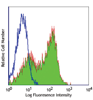
PHA-stimulated (3 days) human peripheral blood lymphocytes were stained with CD25 (clone M-A251) Alexa Fluor® 647 (filled histogram) or mouse IgG1, κ Alexa Fluor® 647 isotype control (open histogram). -

Confocal image of human lymph node sample acquired using the IBEX method of highly multiplexed antibody-based imaging: CD1c (red) in Cycle 3, CD8 (blue) in Cycle 4, and CD25 (yellow) in Cycle 4. Tissues were prepared using ~1% (vol/vol) formaldehyde and a detergent. Following fixation, samples are immersed in 30% (wt/vol) sucrose for cryoprotection. Images are courtesy of Drs. Andrea J. Radtke and Ronald N. Germain of the Center for Advanced Tissue Imaging (CAT-I) in the National Institute of Allergy and Infectious Diseases (NIAID, NIH).
| Cat # | Size | Price | Save |
|---|---|---|---|
| 356127 | 25 tests | ¥27,280 | |
| 356128 | 100 tests | ¥58,300 |
CD25 is a 55 kD type I transmembrane glycoprotein also known as low affinity IL-2 receptor α chain or Tac. It is expressed on progenitor lymphocytes, activated T and B cells, and activated monocytes/macrophages. CD25 is also expressed on a subset of non-stimulated CD4+ T cells termed T regulatory cells. Soluble CD25/IL-2Rα is produced as a consequence of lymphocyte stimulation and is found in biological fluids following inflammatory responses. CD25 associates with IL-2 receptor β (CD122) and common γ (CD132) chains to form a high affinity IL-2R complex.
Product DetailsProduct Details
- Verified Reactivity
- Human
- Reported Reactivity
- Baboon, Cynomolgus, Rhesus
- Antibody Type
- Monoclonal
- Host Species
- Mouse
- Immunogen
- Human PHA-induced lymphocyte cells
- Formulation
- Phosphate-buffered solution, pH 7.2, containing 0.09% sodium azide and BSA (origin USA)
- Preparation
- The antibody was purified by affinity chromatography and conjugated with Alexa Fluor® 647 under optimal conditions.
- Concentration
- Lot-specific (to obtain lot-specific concentration and expiration, please enter the lot number in our Certificate of Analysis online tool.)
- Storage & Handling
- The antibody solution should be stored undiluted between 2°C and 8°C, and protected from prolonged exposure to light. Do not freeze.
- Application
-
FC - Quality tested
SB - Reported in the literature, not verified in house - Recommended Usage
-
Each lot of this antibody is quality control tested by immunofluorescent staining with flow cytometric analysis. For flow cytometric staining, the suggested use of this reagent is 5 µl per million cells in 100 µl staining volume or 5 µl per 100 µl of whole blood.
* Alexa Fluor® 647 has a maximum emission of 668 nm when it is excited at 633 nm / 635 nm.
Alexa Fluor® and Pacific Blue™ are trademarks of Life Technologies Corporation.
View full statement regarding label licenses - Excitation Laser
-
Red Laser (633 nm)
- Application Notes
-
Additional reported applications (for the relevant formats) include: immunohistochemical staining of paraformaldehyde fixed frozen sections1 and spatial biology (IBEX)2,3.
The CD25 molecule reveals three epitope regions: A, B, and C. M-A251 antibody recognizes epitope region B. Unlike other CD25 antibody clones, M-A251 can detect CD25 after fixation with paraformaldehyde. - Additional Product Notes
-
Iterative Bleaching Extended multi-pleXity (IBEX) is a fluorescent imaging technique capable of highly-multiplexed spatial analysis. The method relies on cyclical bleaching of panels of fluorescent antibodies in order to image and analyze many markers over multiple cycles of staining, imaging, and, bleaching. It is a community-developed open-access method developed by the Center for Advanced Tissue Imaging (CAT-I) in the National Institute of Allergy and Infectious Diseases (NIAID, NIH).
-
Application References
(PubMed link indicates BioLegend citation) - Product Citations
-
- RRID
-
AB_2563587 (BioLegend Cat. No. 356127)
AB_2563588 (BioLegend Cat. No. 356128)
Antigen Details
- Structure
- Type I transmembrane glycoprotein, 55 kD; low-affinity IL-2 receptor α chain
- Distribution
-
Activated T and B cells, monocytes/macrophages, Tregs
- Interaction
- Associates with IL-2Rβ/CD122 and IL-2Rγ/CD132 receptor chains to form a high-affinity IL-2R complex
- Ligand/Receptor
- IL-2
- Cell Type
- B cells, Macrophages, Monocytes, T cells, Tregs
- Biology Area
- Cell Biology, Immunology, Neuroscience, Neuroscience Cell Markers
- Molecular Family
- CD Molecules, Cytokine/Chemokine Receptors
- Antigen References
-
1. Knapp W, et al. 1989. Leucocyte Typing IV: White Cell Differentiation Antigens. Oxford University Press.
2. Schlossman S, et al. 1995. Leucocyte Typing V: White Cell Differentiation Antigens. Oxford University Press.
3. Barclay N, et al. 1997. The Leukocyte Antigen FactsBook. Academic Press Inc.
4. Taniguchi T and Minami Y. et al. 1993. Cell 73:5.
5. Waldmann T. 1991. J. Biol. Chem. 266:2681. - Gene ID
- 3559 View all products for this Gene ID
- UniProt
- View information about CD25 on UniProt.org
Related Pages & Pathways
Pathways


Related FAQs
- If an antibody clone has been previously successfully used in IBEX in one fluorescent format, will other antibody formats work as well?
-
It’s likely that other fluorophore conjugates to the same antibody clone will also be compatible with IBEX using the same sample fixation procedure. Ultimately a directly conjugated antibody’s utility in fluorescent imaging and IBEX may be specific to the sample and microscope being used in the experiment. Some antibody clone conjugates may perform better than others due to performance differences in non-specific binding, fluorophore brightness, and other biochemical properties unique to that conjugate.
- Will antibodies my lab is already using for fluorescent or chromogenic IHC work in IBEX?
-
Fundamentally, IBEX as a technique that works much in the same way as single antibody panels or single marker IF/IHC. If you’re already successfully using an antibody clone on a sample of interest, it is likely that clone will have utility in IBEX. It is expected some optimization and testing of different antibody fluorophore conjugates will be required to find a suitable format; however, legacy microscopy techniques like chromogenic IHC on fixed or frozen tissue is an excellent place to start looking for useful antibodies.
- Are other fluorophores compatible with IBEX?
-
Over 18 fluorescent formats have been screened for use in IBEX, however, it is likely that other fluorophores are able to be rapidly bleached in IBEX. If a fluorophore format is already suitable for your imaging platform it can be tested for compatibility in IBEX.
- The same antibody works in one tissue type but not another. What is happening?
-
Differences in tissue properties may impact both the ability of an antibody to bind its target specifically and impact the ability of a specific fluorophore conjugate to overcome the background fluorescent signal in a given tissue. Secondary stains, as well as testing multiple fluorescent conjugates of the same clone, may help to troubleshoot challenging targets or tissues. Using a reference control tissue may also give confidence in the specificity of your staining.
- How can I be sure the staining I’m seeing in my tissue is real?
-
In general, best practices for validating an antibody in traditional chromogenic or fluorescent IHC are applicable to IBEX. Please reference the Nature Methods review on antibody based multiplexed imaging for resources on validating antibodies for IBEX.
Other Formats
View All CD25 Reagents Request Custom ConjugationCustomers Also Purchased
Compare Data Across All Formats
This data display is provided for general comparisons between formats.
Your actual data may vary due to variations in samples, target cells, instruments and their settings, staining conditions, and other factors.
If you need assistance with selecting the best format contact our expert technical support team.
-
APC/Cyanine7 anti-human CD25

PHA-stimulated (3 days) human peripheral blood lymphocytes w... -
Purified anti-human CD25

PHA-stimulated (3 day) human peripheral blood lymphocytes we... -
PE anti-human CD25

PHA-stimulated (3 day) human peripheral blood lymphocytes we... -
FITC anti-human CD25

PHA-stimulated (3 day) human peripheral blood lymphocytes we... -
PE/Cyanine7 anti-human CD25

PHA-stimulated (3 days) human peripheral blood lymphocytes w... -
APC anti-human CD25

PHA-stimulated (3 days) human peripheral blood lymphocytes w... -
PerCP/Cyanine5.5 anti-human CD25

PHA-stimulated (3 days) human peripheral blood lymphocytes w... -
Brilliant Violet 421™ anti-human CD25

PHA-stimulated (3 days) human peripheral blood lymphocytes w... -
Alexa Fluor® 488 anti-human CD25

PHA-stimulated (3 days) human peripheral blood lymphocytes w... -
Alexa Fluor® 700 anti-human CD25

PHA-stimulated (3 days) human peripheral blood lymphocytes w... -
Brilliant Violet 510™ anti-human CD25

PHA-stimulated (3 days) human peripheral blood lymphocytes w... -
PE/Dazzle™ 594 anti-human CD25

PHA-stimulated (3 days) human peripheral blood lymphocytes w... -
Biotin anti-human CD25

PHA-stimulated (3 days) human peripheral blood lymphocytes w... -
Alexa Fluor® 647 anti-human CD25

PHA-stimulated (3 days) human peripheral blood lymphocytes w... 
Confocal image of human lymph node sample acquired using the... -
Pacific Blue™ anti-human CD25

PHA-stimulated (3 days) human peripheral blood lymphocytes w... -
PerCP anti-human CD25

PHA-stimulated (3 days) human peripheral blood lymphocytes w... -
APC/Fire™ 750 anti-human CD25

PHA-stimulated (3 days) human peripheral blood lymphocytes w... -
Brilliant Violet 711™ anti-human CD25

PHA-stimulated (3 days) human peripheral blood lymphocytes w... -
Brilliant Violet 785™ anti-human CD25

PHA-stimulated (3 days) human peripheral blood lymphocytes w... -
Brilliant Violet 605™ anti-human CD25

PHA-stimulated (3 days) human peripheral blood lymphocytes w... 
Human peripheral blood lymphocytes were stained with CD4 APC... -
KIRAVIA Blue 520™ anti-human CD25

PHA-stimulated (3 days) human peripheral blood lymphocytes w... 
Human peripheral blood lymphocytes were stained with CD4 APC... -
PE/Fire™ 700 anti-human CD25

PHA-stimulated (3 days) human peripheral blood lymphocytes w... 
Human peripheral blood lymphocytes were stained with anti-hu... -
APC/Fire™ 810 anti-human CD25

PHA-stimulated (3 days) human peripheral blood lymphocytes w... 
Human peripheral blood lymphocytes were stained with CD4 Bri... -
Spark NIR™ 685 anti-human CD25 Antibody

Human peripheral blood lymphocytes were stained with CD4 FIT... -
Spark YG™ 581 anti-human CD25

PHA-stimulated (3 days) human peripheral blood lymphocytes w... 
Human peripheral blood lymphocytes were stained with anti-hu... -
PE/Fire™ 640 anti-human CD25 Antibody

PHA-stimulated (3 days) human peripheral blood lymphocytes w... 
Human peripheral blood lymphocytes were stained with CD4 Bri... -
PE anti-human CD25

Typical results from human peripheral blood lymphocytes stai... -
PerCP/Cyanine5.5 anti-human CD25

Typical results from human peripheral blood lymphocytes stai... -
APC/Fire™ 750 anti-human CD25

Typical results from human peripheral blood lymphocytes stai... -
PE/Cyanine7 anti-human CD25

Typical results from human peripheral blood lymphocytes stai... -
APC anti-human CD25

Typical results from human peripheral blood lymphocytes stai... -
PE/Cyanine5 anti-human CD25

Human peripheral blood lymphocytes were stained with anti-hu... -
FITC anti-human CD25

Typical results from human peripheral blood lymphocytes stai... -
Spark Red™ 718 anti-human CD25

Human peripheral blood lymphocytes were stained with anti-hu... -
GMP PE anti-human CD25

Typical results from human peripheral blood lymphocytes stai... -
GMP PE/Cyanine7 anti-human CD25

Typical results from human peripheral blood lymphocytes stai... -
PerCP/Fire™ 780 anti-human CD25

Human peripheral blood lymphocytes were stained with anti-hu... -
PE/Fire™ 744 anti-human CD25

PHA-stimulated (3 days) human peripheral blood mononuclear c... 
Human peripheral blood mononuclear cells were stained with a... -
PerCP/Fire™ 806 anti-human CD25

Human peripheral blood lymphocytes were stained with anti-hu... -
Brilliant Violet 650™ anti-human CD25

Human peripheral blood lymphocytes were stained with anti-hu...


Follow Us