


- Clone
- SMI 31 (See other available formats)
- Regulatory Status
- RUO
- Other Names
- Neurofilament heavy polypeptide, NF-H, 200 kD neurofilament protein, neurofilament triplet H protein
- Isotype
- Mouse IgG1, κ
- Ave. Rating
- Submit a Review
- Product Citations
- publications

-

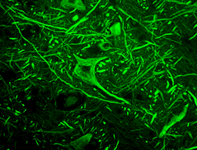
IHC staining of Alexa Fluor® 594 anti-Neurofilament H (NF-H), Phosphorylated (clone SMI 31) on formalin-fixed paraffin-embedded rat brain tissue. Following antigen retrieval using Retrieve-All Antigen Unmasking System 3 (Cat. No. 927701), the tissue was incubated with 5 µg/mL of the primary antibody for 1 hour at room temperature. The image was captured with a 40x objective.
| Cat # | Size | Price | Save |
|---|---|---|---|
| 801609 | 25 µg | ¥28,380 | |
| 801610 | 100 µg | ¥71,060 |
Neurofilaments (NF) are approximately 10 nanometer intermediate filaments found in neurons. They are a major component of the neuronal cytoskeleton, and function primarily to provide structural support for the axon and to regulate the axon diameter. There are three major NF subunits, and the names given to these subunits are based upon the apparent molecular mass of the mammalian subunits on SDS-PAGE. The light or lowest NF (NF-L) runs at 68-70 kD. The medium or middle NF (NF-M) runs at about 145-160 kD, and the heavy or highest NF (NF-H) runs at 200-220 kD. However, the actual molecular weight of these proteins is considerably lower due to the highly charged C-terminal regions of the molecules. The level of NF gene expression correlates with the axonal diameter, which controls how fast electrical signals travel down the axon. Mutant mice with NF abnormalities have phenotypes resembling amyotrophic lateral sclerosis. NF immunostaining is common in diagnostic neuropathology. It is useful for differentiating neurons (positive for NF) from the glia (negative for NF).
Product DetailsProduct Details
- Verified Reactivity
- Human, Mouse, Rat
- Antibody Type
- Monoclonal
- Host Species
- Mouse
- Formulation
- Phosphate-buffered solution, pH 7.2, containing 0.09% sodium azide.
- Preparation
- The antibody was purified by affinity chromatography and conjugated with Alexa Fluor® 594 under optimal conditions.
- Concentration
- 0.5 mg/ml
- Storage & Handling
- The antibody solution should be stored undiluted between 2°C and 8°C, and protected from prolonged exposure to light. Do not freeze.
- Application
-
IHC-P - Quality tested
- Recommended Usage
-
Each lot of this antibody is quality control tested by formalin-fixed paraffin-embedded immunohistochemical staining. For immunohistochemistry, a concentration range of 1.0 - 5.0 µg/ml is suggested. It is recommended that the reagent be titrated for optimal performance for each application.
* Alexa Fluor® 594 has an excitation maximum of 590 nm, and a maximum emission of 617 nm.
Alexa Fluor® and Pacific Blue™ are trademarks of Life Technologies Corporation.
View full statement regarding label licenses - Application Notes
-
Additional reported applications (for the relevant formats) include: Western blotting1, immunohistochemistry2,4, and immunocytochemistry4.
SMI 31 reacts with a phosphorylated epitope in extensively phosphorylated neurofilament H and, to a lesser extent, with neurofilament M in most mammalian species, which chicken and frog (Xenopus). Immunocytochemically, SMI 31 reacts broadly with thick and thin axons and some dendrites such as basket cell dendrites, but not Purkinje cell dendrites. Nerve cell bodies are generally unreactive. Other cells and tissues are unreactive except for peripheral axons. Phosphatase treatment of tissue sections or Western blots abolishes reaction with SMI 31. Staining is unaffected by trypsin. In pathological conditions, reaction with SMI 31 may be found also in neuronal cell bodies. Aberrant phosphorylation of neurofilament H in cell bodies can be demonstrated in neuronal cell cultures with SMI 31 by agents that induce stress-activated protein kinase. In its reaction with paired helical filaments in hereditary inclusion body myopathy, SMI 31 colocalizes with nitric oxide synthase, suggesting that oxidative stress may play a role in the pathogenic cascade of such degenerative diseases. SMI 31 co-immunoprecipitates neurofilament-associated kinase (NAK 115) via reaction of the antibody with the tail domain of neurofilament H. -
Application References
(PubMed link indicates BioLegend citation) - RRID
-
AB_2687300 (BioLegend Cat. No. 801609)
AB_2687298 (BioLegend Cat. No. 801610)
Antigen Details
- Structure
- Neurofilament H has an apparent molecular mass of 200-220 kD.
- Distribution
-
Tissue distribution: CNS, peripheral nerves and glandular cells of the prostate.
Cellular distribution: Cytoskeleton, nucleus, cytosol, and mitochondrion. - Function
- Neurofilaments are the major components of the neuronal cytoskeleton. They provide axonal support and regulate axon diameter.
- Interaction
- Cell bodies and dendrites are generally unstained. Other cells and tissues are unreactive except for peripheral axons.
- Cell Type
- Mature Neurons
- Biology Area
- Cell Biology, Neuroscience, Neuroscience Cell Markers
- Molecular Family
- Intermediate Filaments, Phospho-Proteins
- Antigen References
-
1. Petzold A. 2005. J. Neurol. Sci. 233(1-2):183
- Gene ID
- 4744 View all products for this Gene ID
- UniProt
- View information about Neurofilament H on UniProt.org


Related FAQs
Other Formats
View All Neurofilament H Phospho (NF-H) Reagents Request Custom Conjugation| Description | Clone | Applications |
|---|---|---|
| Purified anti-Neurofilament H (NF-H), Phosphorylated | SMI 31 | IHC-P,WB,ICC |
| Biotin anti-Neurofilament H (NF-H), Phosphorylated | SMI 31 | IHC-P,WB |
| Alexa Fluor® 594 anti-Neurofilament H (NF-H), Phosphorylated | SMI 31 | IHC-P |
| Alexa Fluor® 488 anti-Neurofilament H (NF-H), Phosphorylated | SMI 31 | IHC-P |
| Alexa Fluor® 647 anti-Neurofilament H (NF-H), Phosphorylated | SMI 31 | IHC-P |
| Spark YG™ 570 anti-Neurofilament H (NF-H), Phosphorylated | SMI 31 | IHC-P,IHC-F |
Customers Also Purchased
Compare Data Across All Formats
This data display is provided for general comparisons between formats.
Your actual data may vary due to variations in samples, target cells, instruments and their settings, staining conditions, and other factors.
If you need assistance with selecting the best format contact our expert technical support team.
-
Purified anti-Neurofilament H (NF-H), Phosphorylated

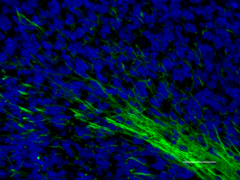
IHC staining of purified anti-Neurofilament H (NF-H), Phosph... 
IHC staining of purified anti-Neurofilament H (NF-H), Phosph... 
Western blot of purified anti-Neurofilament H (NF-H), Phosph... 
IHC staining of purified anti-Neurofilament H (NF-H), Phosph... 
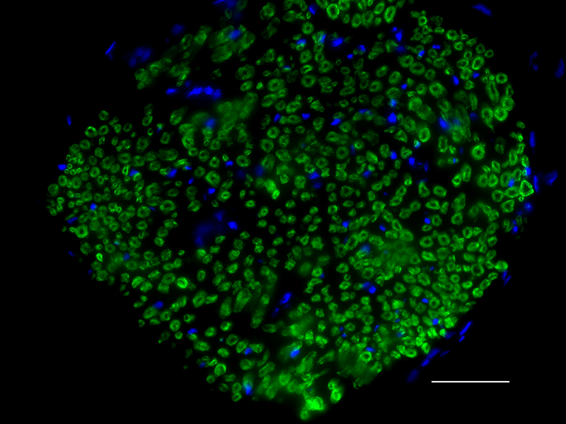
Paraformaldehyde-perfused GFP mouse cerebellum was permeabil... 
ICC staining of purified anti-Neurofilament H (NF-H), Phosph... -
Biotin anti-Neurofilament H (NF-H), Phosphorylated

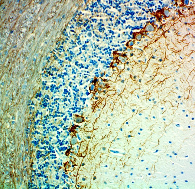
IHC staining of anti-Neurofilament H (NF-H), Phosphorylated ... 
Western blot of anti-Neurofilament H (NF-H), Phosphorylated ... -
Alexa Fluor® 594 anti-Neurofilament H (NF-H), Phosphorylated

IHC staining of Alexa Fluor® 594 anti-Neurofilament H (NF-H... -
Alexa Fluor® 488 anti-Neurofilament H (NF-H), Phosphorylated

IHC staining of Alexa Fluor® 488 anti-Neurofilament H (NF-H)... 
IHC staining of Alexa Fluor® 488 anti-Neurofilament H (NF-H)... -
Alexa Fluor® 647 anti-Neurofilament H (NF-H), Phosphorylated

IHC staining of Alexa Fluor® 647 anti-Neurofilament H (NF-H)... 
IHC staining of Alexa Fluor® 647 anti-Neurofilament H (NF-H)... 
IHC staining of Alexa Fluor® 647 anti-Neurofilament H (NF-H)... 
IHC staining of Alexa Fluor® 647 anti-Neurofilament H (NF-H)... -
Spark YG™ 570 anti-Neurofilament H (NF-H), Phosphorylated

IHC staining of Spark YG™ 570 anti-Neurofilament H (NF-H), P... 
IHC staining of Spark YG™ 570 anti-Neurofilament H (NF-H), P... 
IHC staining of Spark YG™ 570 anti-Neurofilament H (NF-H), P...


Follow Us