


- Clone
- W16155A (See other available formats)
- Regulatory Status
- RUO
- Other Names
- Keratin type II cytoskeletal 7, Cytokeratin-7, CK-7, Keratin-7, K7, Type-II keratin Kb7, Sarcolectin, SCL, Cytokeratin 7, Keratin 7
- Isotype
- Rat IgG2a, κ
- Ave. Rating
- Submit a Review
- Product Citations
- publications

-

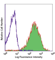
HaCaT cells were fixed with 4% paraformaldehyde (PFA) for 10 minutes, permeabilized with 0.5% Triton X-100 for 3 minutes, and blocked with 5% FBS for 60 minutes. Then the cells were intracellularly stained overnight at 4°C with (A) 1: 100 diluted (5 µg/ml) Alexa Fluor® 488 rat IgG2a (Cat. No. 400525) or with Alexa Fluor® 488 anti-human Cytokeratin 7 antibody (green) at (B) 1:100 (5 µg/ml) and (C) 1:200 (2.5 µg/ml) dilution. Nuclei were counterstained with DAPI (blue). The image was captured with a 60X objective. -

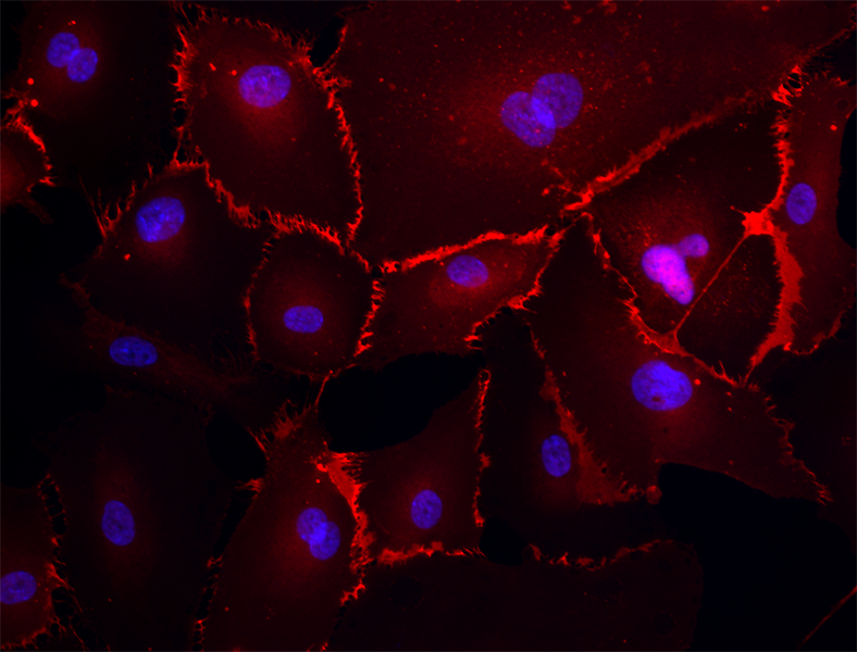
Confocal image of human liver sample acquired using the IBEX method of highly multiplexed antibody-based imaging: Cytokeratin 7 (green) in Cycle 1, CD44 (blue) in Cycle 4, and CD45 (magenta) in Cycle 4. Tissues were prepared using ~1% (vol/vol) formaldehyde and a detergent. Following fixation, samples are immersed in 30% (wt/vol) sucrose for cryoprotection. Images are courtesy of Drs. Andrea J. Radtke and Ronald N. Germain of the Center for Advanced Tissue Imaging (CAT-I) in the National Institute of Allergy and Infectious Diseases (NIAID, NIH).
| Cat # | Size | Price | Save |
|---|---|---|---|
| 601605 | 25 µg | ¥34,980 | |
| 601606 | 100 µg | ¥81,400 |
Keratin 7 (KRT7) belongs to type II keratin and is expressed in the epithelia lining the cavities of internal organs, gland ducts and blood vessels. The function of KRT7 is still not clear but it has been suggested involved in the translational regulation of the human papillomavirus type 16 E7 mRNA (HPV16 E7). In cancer, the expression of KRT7 is observed in many types of carcinoma and is associated with neoplasm progression. The diverse and unique expression pattern of KRT7 is often used as a diagnosis indicator in epithelial origin carcinoma. For example, aberrant KRT7 expression is observed in triple negative breast cancer. In bladder cancer, loss of KRT7 results in promoting proliferation of the bladder urothelium.
Product DetailsProduct Details
- Verified Reactivity
- Human
- Antibody Type
- Monoclonal
- Host Species
- Rat
- Immunogen
- Human KRT7 peptide (446-462) conjugated to KLH.
- Formulation
- Phosphate-buffered solution, pH 7.2, containing 0.09% sodium azide.
- Preparation
- The antibody was purified by affinity chromatography and conjugated with Alexa Fluor® 488 under optimal conditions.
- Concentration
- 0.5 mg/ml
- Storage & Handling
- The antibody solution should be stored undiluted between 2°C and 8°C, and protected from prolonged exposure to light. Do not freeze.
- Application
-
ICC - Quality tested
SB - Reported in the literature, not verified in house - Recommended Usage
-
Each lot of this antibody is quality control tested by immunocytochemistry. For immunocytochemistry, a concentration range of 2.5 - 5.0 μg/ml (1:100-1:200 dilution) is recommended. It is recommended that the reagent be titrated for optimal performance for each application.
* Alexa Fluor® 488 has a maximum emission of 519 nm when it is excited at 488 nm.
Alexa Fluor® and Pacific Blue™ are trademarks of Life Technologies Corporation.
View full statement regarding label licenses - Excitation Laser
-
Blue Laser (488 nm)
- Application Notes
-
This antibody does not react with mouse (in-house tested). Additional reported applications (for the relevant formats) include: spatial biology (IBEX)1,2.
- Additional Product Notes
-
Iterative Bleaching Extended multi-pleXity (IBEX) is a fluorescent imaging technique capable of highly-multiplexed spatial analysis. The method relies on cyclical bleaching of panels of fluorescent antibodies in order to image and analyze many markers over multiple cycles of staining, imaging, and, bleaching. It is a community-developed open-access method developed by the Center for Advanced Tissue Imaging (CAT-I) in the National Institute of Allergy and Infectious Diseases (NIAID, NIH).
-
Application References
(PubMed link indicates BioLegend citation) - RRID
-
AB_2728458 (BioLegend Cat. No. 601605)
AB_2728459 (BioLegend Cat. No. 601606)
Antigen Details
- Structure
- Type II cytokeratin. 469 amino acids with a predicted molecular weight of 51 kD.
- Distribution
-
Cytoplasm.
- Function
- KRT7 is a type II keratin, involved in the translational regulation of the human papillomavirus type 16 E7 mRNA.
- Interaction
- KRT7 interacts with EIF3S10, HPV16 E7, and GPER1.
- Biology Area
- Cell Biology, Cell Motility/Cytoskeleton/Structure, Neuroscience, Neuroscience Cell Markers
- Molecular Family
- Intermediate Filaments
- Antigen References
-
1. Tajima Y, et al. 2017. Sci Rep. 7:40684.
2. Huang B, et al. 2016. Oncogene. 35:4927.
3. Szponar A, et al. 2012. Virchows Arch. 460:423.
4. Sano M, et al. 2010. Int J Oncol. 36:321. - Gene ID
- 3855 View all products for this Gene ID
- UniProt
- View information about Cytokeratin 7 on UniProt.org


Related FAQs
- If an antibody clone has been previously successfully used in IBEX in one fluorescent format, will other antibody formats work as well?
-
It’s likely that other fluorophore conjugates to the same antibody clone will also be compatible with IBEX using the same sample fixation procedure. Ultimately a directly conjugated antibody’s utility in fluorescent imaging and IBEX may be specific to the sample and microscope being used in the experiment. Some antibody clone conjugates may perform better than others due to performance differences in non-specific binding, fluorophore brightness, and other biochemical properties unique to that conjugate.
- Will antibodies my lab is already using for fluorescent or chromogenic IHC work in IBEX?
-
Fundamentally, IBEX as a technique that works much in the same way as single antibody panels or single marker IF/IHC. If you’re already successfully using an antibody clone on a sample of interest, it is likely that clone will have utility in IBEX. It is expected some optimization and testing of different antibody fluorophore conjugates will be required to find a suitable format; however, legacy microscopy techniques like chromogenic IHC on fixed or frozen tissue is an excellent place to start looking for useful antibodies.
- Are other fluorophores compatible with IBEX?
-
Over 18 fluorescent formats have been screened for use in IBEX, however, it is likely that other fluorophores are able to be rapidly bleached in IBEX. If a fluorophore format is already suitable for your imaging platform it can be tested for compatibility in IBEX.
- The same antibody works in one tissue type but not another. What is happening?
-
Differences in tissue properties may impact both the ability of an antibody to bind its target specifically and impact the ability of a specific fluorophore conjugate to overcome the background fluorescent signal in a given tissue. Secondary stains, as well as testing multiple fluorescent conjugates of the same clone, may help to troubleshoot challenging targets or tissues. Using a reference control tissue may also give confidence in the specificity of your staining.
- How can I be sure the staining I’m seeing in my tissue is real?
-
In general, best practices for validating an antibody in traditional chromogenic or fluorescent IHC are applicable to IBEX. Please reference the Nature Methods review on antibody based multiplexed imaging for resources on validating antibodies for IBEX.
Other Formats
View All Cytokeratin 7 Reagents Request Custom Conjugation| Description | Clone | Applications |
|---|---|---|
| Purified anti-human Cytokeratin 7 | W16155A | WB,ICC |
| Alexa Fluor® 594 anti-human Cytokeratin 7 | W16155A | ICC |
| Alexa Fluor® 488 anti-human Cytokeratin 7 | W16155A | ICC,SB |
| Alexa Fluor® 647 anti-human Cytokeratin 7 | W16155A | ICC |
Customers Also Purchased
Compare Data Across All Formats
This data display is provided for general comparisons between formats.
Your actual data may vary due to variations in samples, target cells, instruments and their settings, staining conditions, and other factors.
If you need assistance with selecting the best format contact our expert technical support team.
-
Purified anti-human Cytokeratin 7

Total cell lysate (15ug protein) from HaCaT, A549, MCF-7 (ne... 
HaCaT cells were fixed with 4% paraformaldehyde (PFA) for 1... 
HaCaT cells were fixed with 4% paraformaldehyde (PFA) for 15... -
Alexa Fluor® 594 anti-human Cytokeratin 7

HaCaT cells were fixed with 4% paraformaldehyde (PFA) for 1... 
HaCaT cells were fixed with 4% paraformaldehyde (PFA) for 1... -
Alexa Fluor® 488 anti-human Cytokeratin 7

HaCaT cells were fixed with 4% paraformaldehyde (PFA) for 10... 
Confocal image of human liver sample acquired using the IBEX... -
Alexa Fluor® 647 anti-human Cytokeratin 7

HaCaT cells were fixed with 4% paraformaldehyde (PFA) for 10...


Follow Us