 Login / Register


- Clone
- DO-1 (See other available formats)
- Regulatory Status
- RUO
- Other Names
- Tumor protein p53 (TP53)
- Isotype
- Mouse IgG2a, κ
- Ave. Rating
- Submit a Review
- Product Citations
- publications

-

A431 cells were fixed with 4% paraformaldehyde for 10 minutes, permeabilized with Triton X-100 for 10 minutes, and blocked with 5% FBS for 60 minutes. Cells were then intracellularly stained with a 1:250 dilution (2 µg/mL) of either Alexa Fluor® 594 Mouse IgG2a, κ Isotype Control Antibody (Cat. No. 400280, panel A) or Alexa Fluor® 594 anti-p53 Antibody (panel B) for 2 hours at room temperature. Nuclei were counterstained with DAPI and the image was captured with a 60X objective.
| Cat # | Size | Price | Quantity Check Availability | Save | ||
|---|---|---|---|---|---|---|
| 645705 | 25 µg | 113€ | ||||
| 645706 | 100 µg | 259€ | ||||
p53 is a 53 kD protein that forms tetramers and functions as a tumor suppressor and transcriptional activator of genes that inhibit growth and/or invasion, cell cycle checkpoint after irradiation, DNA repair, apoptotic induction, signal transduction, and cell adhesion. This protein is localized to the nucleus when activated and can be upregulated by genotoxic or other cellular stresses. p53 is modified by phosphorylation, acetylation, ribosylation, ubiquitination, and sumoylation; ubiquination targets p53 for degradation via mdm2. This protein interacts with a variety of proteins including mdm2, mdmx, topoisomerase I, PML3, Bcl-XL, Bcl-2, Chk1, JNK, p38, HIPK2, CK2, DNA-PK, p300/CBP, PCAF, PARP1, and HDAC1-3. Mutant p53 associates with p63 and p73.
Product DetailsProduct Details
- Verified Reactivity
- Human
- Antibody Type
- Monoclonal
- Host Species
- Mouse
- Immunogen
- P53 N-terminal linear epitope aa 20-25
- Formulation
- Phosphate-buffered solution, pH 7.2, containing 0.09% sodium azide.
- Preparation
- The antibody was purified by affinity chromatography and conjugated with Alexa Fluor® 594 under optimal conditions.
- Concentration
- 0.5 mg/mL
- Storage & Handling
- The antibody solution should be stored undiluted between 2°C and 8°C, and protected from prolonged exposure to light. Do not freeze.
- Application
-
ICC - Quality tested
- Recommended Usage
-
Each lot of this antibody is quality control tested by immunocytochemistry. For immunocytochemistry, a concentration range of 0.5 - 2.0 μg/mL is recommended. It is recommended that the reagent be titrated for optimal performance for each application.
* Alexa Fluor® 594 has an excitation maximum of 590 nm, and a maximum emission of 617 nm.
Alexa Fluor® and Pacific Blue™ are trademarks of Life Technologies Corporation.
View full statement regarding label licenses - Application References
-
- Vojtesek B, et al. 1992. J Immuno Methods 151:237
- Stephen CW, et al. 1995. J Mol Biol 248:58
- RRID
-
AB_2814494 (BioLegend Cat. No. 645705)
AB_2814495 (BioLegend Cat. No. 645706)
Antigen Details
- Structure
- Tetramer; 53 kD
- Distribution
-
Nuclear when activated
- Function
- Tumor suppressor, transcription factor regulates cell cycle checkpoint, apoptosis, DNA integrity
- Interaction
- mdm2, mdmx, TopoI, PML3, Bcl-XL, Bcl-2, Chk1, JNK, p38, HIPK2, CK2, DNA-PK, p300/CBP, PCAF, PARP1, HDAC1-3; mutant p53 associates with p63, p73
- Cell Type
- Mesenchymal Stem Cells
- Biology Area
- Cell Biology, DNA Repair/Replication, Stem Cells, Transcription Factors
- Antigen References
-
1. Vogelstein B, et al. 1992. Cell. 70:523.
2. Shieh S, et al. 1997. Cell. 91:325.
3. Mihara M, et al. 2003. Mol. Cell 11:577.
4. Saito S, et al. 2003. J. Biol. Chem. 278:37536. - Gene ID
- 7157 View all products for this Gene ID
- UniProt
- View information about p53 on UniProt.org
Related Pages & Pathways
Pathways


Related FAQs
Other Formats
View All p53 Reagents Request Custom Conjugation| Description | Clone | Applications |
|---|---|---|
| Purified anti-p53 | DO-1 | WB,ICC,IP,ChIP |
| Alexa Fluor® 594 anti-p53 | DO-1 | ICC |
Customers Also Purchased
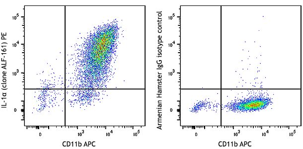
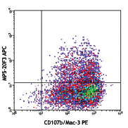
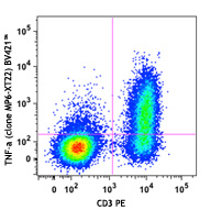
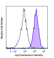
Compare Data Across All Formats
This data display is provided for general comparisons between formats.
Your actual data may vary due to variations in samples, target cells, instruments and their settings, staining conditions, and other factors.
If you need assistance with selecting the best format contact our expert technical support team.


Follow Us