 Login / Register


- Clone
- SK1 (See other available formats)
- Regulatory Status
- RUO
- Other Names
- T8, Leu2
- Isotype
- Mouse IgG1, κ
- Ave. Rating
- Submit a Review
- Product Citations
- publications

-

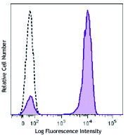
Human peripheral blood lymphocytes were stained with CD8 (clone SK1) Spark Blue™ 550 (filled histogram.) Open histogram represents unstained cells.
| Cat # | Size | Price | Quantity Check Availability | Save | ||
|---|---|---|---|---|---|---|
| 344759 | 25 tests | 113€ | ||||
| 344760 | 100 tests | 296€ | ||||
CD8a is a 32-34 kD type I glycoprotein. It forms a homodimer (CD8a/a) or heterodimer (CD8a/b) with CD8b. CD8, also known as T8 and Leu2, is a member of the immunoglobulin superfamily found on the majority of thymocytes, a subset of peripheral blood T cells, and NK cells (which express almost exclusively CD8a homodimers). CD8 acts as a co-receptor with MHC class I-restricted T cell receptors in antigen recognition and T cell activation and has been shown to play a role in thymic differentiation. Two domains in CD8a are important for function: the extracellular IgSF domain binds the α3 domain of MHC class I and the cytoplasmic CXCP motif binds the tyrosine kinase p56 Lck.
Product DetailsProduct Details
- Verified Reactivity
- Human, Cynomolgus, Rhesus
- Reported Reactivity
- African Green, Chimpanzee, Pigtailed Macaque, Sooty Mangabey
- Antibody Type
- Monoclonal
- Host Species
- Mouse
- Formulation
- Phosphate-buffered solution, pH 7.2, containing 0.09% sodium azide and BSA (origin USA)
- Preparation
- The antibody was purified by affinity chromatography and conjugated with Spark Blue™ 550 under optimal conditions.
- Concentration
- Lot-specific (to obtain lot-specific concentration and expiration, please enter the lot number in our Certificate of Analysis online tool.)
- Storage & Handling
- The antibody solution should be stored undiluted between 2°C and 8°C, and protected from prolonged exposure to light. Do not freeze.
- Application
-
FC - Quality tested
- Recommended Usage
-
Each lot of this antibody is quality control tested by immunofluorescent staining with flow cytometric analysis. For flow cytometric staining, the suggested use of this reagent is 5 µL per million cells in 100 µL staining volume or 5 µL per 100 µL of whole blood. It is recommended that the reagent be titrated for optimal performance for each application.
* Spark Blue™ 550 has a maximum excitation of 516 nm and a maximum emission of 540 nm. - Excitation Laser
-
Blue Laser (488 nm)
- Application Notes
-
Clone SK1 recognizes the a chain of CD8. Additional reported applications (for the relevant formats) include: proteogenomics8, immunohistochemistry of acetone-fixed frozen tissue sections, and spatial biology (IBEX)9,10. This clone was tested in-house and does not demonstrate utility for formalin-fixed paraffin-embedded (FFPE) human tonsil sections.
- Application References
-
- Ledbetter JA, et al. 1981. J. Exp. Med. 153:310.
- Campanelli R, et al. 2002. Intl. Immunol. 14:39.
- Evans RL, et al. 1981. Immunol. 78:544.
- Wooldridge L, et al. 2005. J. Bio. Chem. 280:27491.
- Ch'el IL, et al. 2011. J Exp Med. 208:633. PubMed
- Carbone A, et al. 1999. Blood 93:2319. (IHC-F)
- Ahmed A, et al. 2001. J. Pathol. 193:383. (IHC)
- Peterson VM, et al. 2017. Nat. Biotechnol. 35:936. (PG)
- Radtke AJ, et al. 2020. Proc Natl Acad Sci USA. 117:33455-33465. (SB) PubMed
- Radtke AJ, et al. 2022. Nat Protoc. 17:378-401. (SB) PubMed
- Product Citations
-
- RRID
-
AB_2819982 (BioLegend Cat. No. 344759)
AB_2819983 (BioLegend Cat. No. 344760)
Antigen Details
- Structure
- Ig superfamily, homodimer or heterodimer with CD8b, 32-34 kD
- Distribution
-
Majority of thymocytes, T cell subset, NK cells
- Function
- MHC class I co-receptor, thymic differentiation, T cell activation
- Ligand/Receptor
- MHC Class I molecules
- Cell Type
- NK cells, T cells, Thymocytes
- Biology Area
- Immunology
- Molecular Family
- CD Molecules
- Antigen References
-
1. Barclay N, et al. 1993. The Leucocyte Antigen FactsBook. Academic Press Inc. San Diego.
- Gene ID
- 925 View all products for this Gene ID
- UniProt
- View information about CD8 on UniProt.org
Related Pages & Pathways
Pathways


Related FAQs
Other Formats
View All CD8 Reagents Request Custom ConjugationCustomers Also Purchased
Compare Data Across All Formats
This data display is provided for general comparisons between formats.
Your actual data may vary due to variations in samples, target cells, instruments and their settings, staining conditions, and other factors.
If you need assistance with selecting the best format contact our expert technical support team.
-
Alexa Fluor® 647 anti-human CD8

Human peripheral blood lymphocytes were stained with CD8 (cl... -
Brilliant Violet 650™ anti-human CD8

Human peripheral blood lymphocytes were stained with CD8 (cl... -
Purified anti-human CD8

Human peripheral blood lymphocytes stained with SK1, followe... -
FITC anti-human CD8
Human peripheral blood lymphocytes stained with SK1 FITC -
PE anti-human CD8

Human peripheral blood lymphocytes stained with SK1 PE. -
PerCP anti-human CD8

Human peripheral blood lymphocytes stained with SK1 PerCP -
PerCP/Cyanine5.5 anti-human CD8

Human peripheral blood lymphocytes stained with CD8 (clone S... -
PE/Cyanine7 anti-human CD8

Human peripheral blood lymphocytes stained with SK1 PE/Cyani... -
APC/Cyanine7 anti-human CD8

Human peripheral blood lymphocytes were stained with CD8 (cl... -
Alexa Fluor® 488 anti-human CD8

Human peripheral blood lymphocytes stained with SK1 Alexa Fl... 
Confocal image of human lymph node sample acquired using the... 
Confocal image of human metastatic lymph node sample acquire... -
Pacific Blue™ anti-human CD8

Human peripheral blood lymphocytes stained with SK1 Pacific ... -
Biotin anti-human CD8

Human peripheral blood lymphocytes stained with SK1 biotin, ... -
APC anti-human CD8

Human peripheral blood lymphocytes stained with SK1 APC -
Alexa Fluor® 700 anti-human CD8

Human peripheral blood lymphocytes were stained with CD8 (cl... -
Purified anti-human CD8 (Maxpar® Ready)

Human PBMCs stained with 154Sm-anti-CD45 (HI30) and 151Eu-an... -
Brilliant Violet 510™ anti-human CD8

Human peripheral blood lymphocytes were stained with CD8 (cl... -
Brilliant Violet 711™ anti-human CD8

Human peripheral blood lymphocytes were stained with CD8 (cl... -
Brilliant Violet 785™ anti-human CD8

Human peripheral blood lymphocytes were stained with CD8 (cl... -
Brilliant Violet 605™ anti-human CD8

Human peripheral blood lymphocytes were stained with CD8 (cl... -
PE/Dazzle™ 594 anti-human CD8

Human peripheral blood lymphocytes were stained with CD8 (cl... -
PE anti-human CD8

Typical results from human peripheral blood lymphocytes stai... -
APC/Fire™ 750 anti-human CD8

Human peripheral blood lymphocytes were stained with CD3 PE ... 
-
APC anti-human CD8

Typical results from human peripheral blood lymphocytes stai... -
Brilliant Violet 421™ anti-human CD8

Human peripheral blood lymphocytes were stained with CD8 (cl... -
Pacific Blue™ anti-human CD8

Typical results from human peripheral blood lymphocytes stai... -
TotalSeq™-A0046 anti-human CD8
-
TotalSeq™-C0046 anti-human CD8
-
Brilliant Violet 750™ anti-human CD8

Human peripheral blood lymphocytes were stained with CD8 (cl... -
TotalSeq™-B0046 anti-human CD8
-
Spark Blue™ 550 anti-human CD8

Human peripheral blood lymphocytes were stained with CD8 (cl... -
PE/Cyanine7 anti-human CD8
Typical results from human peripheral blood lymphocytes stai... -
FITC anti-human CD8
Typical results from human peripheral blood lymphocytes stai... -
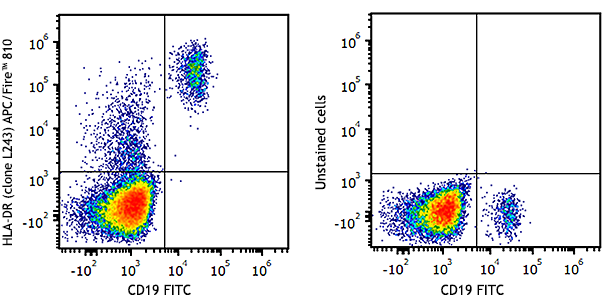
APC/Fire™ 810 anti-human CD8

Human peripheral blood lymphocytes were stained with CD8 (cl... -
PE/Fire™ 640 anti-human CD8

Human peripheral blood lymphocytes were stained with CD8 (cl... -
PE/Fire™ 700 anti-human CD8

Human peripheral blood lymphocytes were stained with anti-hu... -
PerCP anti-human CD8

Typical results from human peripheral blood lymphocytes stai... -
PerCP/Cyanine5.5 anti-human CD8

Typical results from human peripheral blood lymphocytes stai... -
TotalSeq™-D0046 anti-human CD8
-
APC/Fire™ 750 anti-human CD8

Typical results from human peripheral blood lymphocytes stai... -
GMP APC anti-human CD8

Typical results from human peripheral blood lymphocytes stai... -
PE/Cyanine5 anti-human CD8 Antibody

Human peripheral blood lymphocytes were stained with anti-hu... -
Spark UV™ 387 anti-human CD8

Human peripheral blood lymphocytes were stained with anti-hu... -
GMP PE anti-human CD8

Typical results from human peripheral blood lymphocytes stai... -
GMP PE/Cyanine7 anti-human CD8

Typical results from human peripheral blood lymphocytes stai... -
Spark NIR™ 685 anti-human CD8

Human peripheral blood lymphocytes were stained with anti-hu... -
KIRAVIA Blue 520™ anti-human CD8

Human peripheral blood lymphocytes stained with anti-human C... -
GMP FITC anti-human CD8

Typical results from human peripheral blood lymphocytes stai... -
GMP Pacific Blue™ anti-human CD8

Typical results from human peripheral blood lymphocytes stai... -
GMP PerCP anti-human CD8

Typical results from human peripheral blood lymphocytes stai... -
Spark Violet™ 500 anti-human CD8

Human peripheral blood lymphocytes were stained with anti-hu... -
GMP APC/Fire™ 750 anti-human CD8

Typical results from human peripheral blood lymphocytes stai... -
GMP PerCP/Cyanine 5.5 anti-human CD8

Typical results from human peripheral blood lymphocytes stai... -
Alexa Fluor® 660 anti-human CD8a

Human peripheral blood lymphocytes were stained with anti-hu... -
Spark Blue™ 515 anti-human CD8

Human peripheral blood lymphocytes were stained with anti-hu... -
Spark Blue™ 574 anti-human CD8

Human peripheral blood lymphocytes were stained with anti-hu... -
Spark Violet™ 538 anti-human CD8

Human peripheral blood lymphocytes were stained with anti-hu... -
PE/Fire™ 810 anti-human CD8

Human peripheral blood lymphocytes were stained with anti-hu...


Follow Us