 Login / Register


- Clone
- 5A5 (See other available formats)
- Regulatory Status
- RUO
- Other Names
- Poly (ADP-ribose) polymerase
- Isotype
- Mouse IgG1, κ
- Ave. Rating
- Submit a Review
- Product Citations
- publications

-

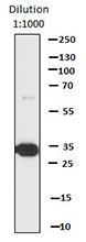
Western blot analysis of cell lysates from HeLa cells, untreated (-) or treated with Staurosporine (+, 1 µM, 8 hours), using PARP (5A5) Mouse primary antibody and HRP Goat anti-Mouse secondary antibody (#405306). Direct-Blot™ HRP anti-β-actin (#643807) was used as a loading control. -

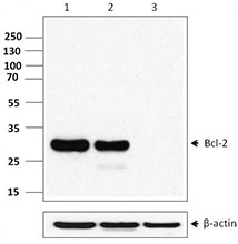
Total lysates (15 µg protein) from 293T and 293T/PARP knockdown (KD) cells were resolved by electrophoresis (4-20% Tris-Glycine gel), transferred to nitrocellulose, and probed with 1:1000 diluted (0.5 µg/mL) purified anti-PARP antibody, clone 5A5 (upper). Proteins were visualized using chemiluminescence detection by incubating with 1:3000 dilution of HRP goat anti-mouse-IgG secondary antibody (Cat. No. 405306). 1:2000 dilution of Direct-Blot™ HRP anti-β-actin Antibody (Cat. No. 643807) was used as a loading control (lower). Lane M: MW ladder. -

HeLa cells were fixed with 1% paraformaldehyde (PFA) for 10 minutes, permeabilized with 0.5% Triton X-100 for 10 minutes and blocked with 5% FBS for 30 minutes. Then the cells were intracellularly stained with 5 µg/ml of purified PARP (clone 5A5) (red) in blocking buffer overnight at 4°C and followed by anti-mouse IgG Dylight™ 594 staining for 2 hours and Alexa Fluor® 488 Phalloidin (green) staining for 20 minutes at 4°C . Nuclei were counterstained with DAPI (blue). The image was captured with 40X objective. -

IHC staining using purified anti-PARP antibody (clone 5A5) on formalin-fixed paraffin-embedded tonsil tissue. The tissue was incubated with 10 µg/mL of antibody overnight at 4°C, followed by incubation with 2.5 µg/mL of Alexa Fluor® 647 goat anti-mouse IgG antibody (red, both panels) (Cat. No. 405322) for one hour at room temperature. Nuclei were counterstained with DAPI (blue, left panel) (Cat. No. 422801), and the slide was mounted with ProLong™ Gold Antifade Mountant. The image was captured with a 40x objective. Scalebar = 50 µM
| Cat # | Size | Price | Quantity Check Availability | Save | ||
|---|---|---|---|---|---|---|
| 614301 | 25 µg | 81€ | ||||
| 614302 | 100 µg | 177€ | ||||
PARP (Poly (ADP-ribose) polymerase) is a 113 kD nuclear protein that can exist as a homo- or hetero-dimer. This protein acts as a molecular "nick sensor" and functions in base excision repair, poly(ADP-ribosyl)ation of acceptor proteins involved in chromatin architecture and DNA metabolism and participates in protein modification to enhance or repress transcription. PARP is ribosylated by PARP2 and is a target for caspase cleavage during apoptosis. PARP interacts with proteins in the base excision repair complex containing at least XRCC1, PARP2, POLB and LIG3. In addition PARP forms heterodimers with PARP2, and interacts with PARP3. The 5A5 monoclonal antibody recognizes the N-terminal region of human and mouse PARP and has been shown to be useful for Western blotting and immunofluorescence staining.
Product DetailsProduct Details
- Verified Reactivity
- Human
- Antibody Type
- Monoclonal
- Host Species
- Mouse
- Immunogen
- Recombinant (partial), N-terminal 2/3 sequence of PARP
- Formulation
- This antibody is provided in phosphate-buffered solution, pH 7.2, containing 0.09% sodium azide. Final antibody concentration is 0.5 mg/ml.
- Preparation
- The antibody was purified by affinity chromatography.
- Concentration
- 0.5 mg/ml
- Storage & Handling
- Upon receipt, store undiluted between 2°C and 8°C.
- Application
-
WB - Quality tested
KO/KD-WB, ICC, IHC-P - Verified - Recommended Usage
-
Each lot of this antibody is quality control tested by Western blotting. For Western blotting, the suggested use of this reagent is 0.5 - 1.0 µg/mL (1:500-1:1000 dilution). For immunocytochemistry, a concentration range of 1 - 5 μg/mL is recommended. For immunohistochemistry, a concentration range of 5 - 10 µg/mL is suggested. It is recommended that the reagent be titrated for optimal performance for each application.
- Application Notes
-
This antibody recognizes N-terminal short cleaved PARP after Staurosporine treatment at around 24kDa.
- Application References
- Product Citations
-
- RRID
-
AB_2160595 (BioLegend Cat. No. 614301)
AB_2299318 (BioLegend Cat. No. 614302)
Antigen Details
- Structure
- PARP family, BRCT domain, homo- or hetero-dimer; 113 kD
- Distribution
-
Nuclear
- Function
- Molecular "nick sensor"; base excision repair, catalyzes poly(ADP-ribosyl)ation of acceptor proteins involved in chromatin architecture, DNA metabolism; protein modification may enhance or repress transcription
- Interaction
- Component of a base excision repair complex containing at least XRCC1, PARP2, POLB and LIG3. Heterodimerizes with PARP2, interacts with PARP3, modifies TATA-BP, YY1, Sp1, NF-B, p53 and others
- Modification
- Ribosylation by PARP2
- Biology Area
- Apoptosis/Tumor Suppressors/Cell Death, Cell Biology, DNA Repair/Replication, Transcription Factors
- Molecular Family
- Nuclear Markers
- Antigen References
-
1. Cherney B, et al. 1987. P. Natl. Acad. Sci. USA 84:8370.
2. Ikejima M, et al. 1990. J. Biol. Chem. 265:21907.
3. Ying W, et al. 2001. P. Natl. Acad. Sci. USA 98:12227.
4. Noel G, et al. 2003. BMC Cell Biol. 4:7. - Regulation
- Poly(ADP-ribose) glycohydrolase removes ribose chains, allows quick, transient ribosylation of proteins
- Gene ID
- 142 View all products for this Gene ID
- UniProt
- View information about PARP on UniProt.org

Related FAQs
Other Formats
View All PARP Reagents Request Custom Conjugation| Description | Clone | Applications |
|---|---|---|
| Purified anti-PARP | 5A5 | WB,KO/KD-WB,ICC,IHC-P |
| Alexa Fluor® 594 anti-PARP | 5A5 | ICC,IHC-P |
| Alexa Fluor® 647 anti-PARP | 5A5 | ICC,IHC-P |
Customers Also Purchased
Compare Data Across All Formats
This data display is provided for general comparisons between formats.
Your actual data may vary due to variations in samples, target cells, instruments and their settings, staining conditions, and other factors.
If you need assistance with selecting the best format contact our expert technical support team.
-
Purified anti-PARP

Western blot analysis of cell lysates from HeLa cells, untre... 
Total lysates (15 µg protein) from 293T and 293T/PARP knockd... 
HeLa cells were fixed with 1% paraformaldehyde (PFA) for 10 ... 
IHC staining using purified anti-PARP antibody (clone 5A5) o... -
Alexa Fluor® 594 anti-PARP

HeLa cells were fixed with 1% paraformaldehyde (PFA) for 10 ... 
Human paraffin-embedded tonsil tissue slices were prepared w... 
Human paraffin-embedded Breast Cancer tissue slices were pre... 
Human paraffin-embedded Breast Cancer tissue slices were pre... -
Alexa Fluor® 647 anti-PARP

HeLa cells were fixed with 1% paraformaldehyde (PFA) for 10 ... 
Human paraffin-embedded tonsil tissue slices were prepared w... 
Human paraffin-embedded Breast Cancer tissue slices were pre...


Follow Us