 Login / Register


- Clone
- 1B5.H9 (See other available formats)
- Regulatory Status
- RUO
- Other Names
- P60, p62, A170, OSIL, PDB3, ZIP3, p62B, sequestosome-1, oxidative stress-induced-like ubiquitin-binding protein p62
- Previously
-
Covance Catalog# MMS-5034
- Isotype
- Mouse IgG1, κ
- Ave. Rating
- Submit a Review
- Product Citations
- publications

-

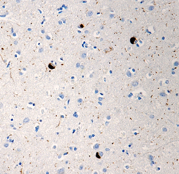
IHC staining of purified anti-p62 (SQSTM1) antibody (clone 1B5.H9) on formalin-fixed paraffin-embedded mouse brain tissue. Following antigen retrieval using Sodium Citrate H.I.E.R., the tissue was incubated with 15 µg/ml of the primary antibody overnight at 4°C. BioLegend´s Ultra-Streptavidin (USA) HRP kit (Multi-Species, DAB, Cat. No. 929901) was used for detection followed by hematoxylin counterstaining, according to the protocol provided. The image was captured with a 40X objective. Scale bar: 50 µm -

IHC staining of purified anti-p62 (SQSTM1) antibody (clone 1B5.H9) on formalin-fixed paraffin-embedded rat brain tissue. Following antigen retrieval using Sodium Citrate H.I.E.R., the tissue was incubated with 15 µg/ml of the primary antibody overnight at 4°C. BioLegend´s Ultra-Streptavidin (USA) HRP kit (Multi-Species, DAB, Cat. No. 929901) was used for detection followed by hematoxylin counterstaining, according to the protocol provided. The image was captured with a 40X objective. Scale bar: 50 µm -

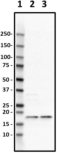
Western blot of purified anti-p62 (SQSTM1) antibody (clone 1B5.H9). Lane 1: Molecular weight marker; Lane 2: 20 µg of HEK293 cell lysate; Lane 3: 20 µg of SH-SY5Y cell lysate. The blot was incubated with 5 µg/mL of the primary antibody overnight at 4°C, followed by incubation with HRP labeled goat anti-mouse IgG (Cat. No. 405306). Enhanced chemiluminescence was used as the detection system. -

Whole cell extracts (15 µg protein) from HeLa cells untreated (-) or treated (+) with 50 µM chloroquine phosphate (Cat. No. 427502) for 18 hours were resolved by 4-12% Bis-Tris gel electrophoresis, transferred to a PVDF membrane, and probed with 1.0 µg/mL purified anti-p62 (SQSTM1) antibody (clone 1B5.H9) overnight at 4°C. Proteins were visualized by chemiluminescence detection using HRP donkey anti-mouse IgG antibody (Cat. No. 405306) at a 1:3000 dilution. Western-Ready™ ECL Substrate Premium Kit (Cat. No. 426319) was used as a detection agent. Direct-Blot™ HRP anti-GAPDH antibody (Cat. No. 607904) was used as a loading control at a 1:25000 dilution (lower). Lane M: Molecular weight marker. -

ICC staining of purified anti-p62 (SQSTM1) antibody (clone 1B5.H9) on A549 cells. The cells were fixed with 4% PFA, permeabilized with a buffer containing 0.1% Triton X-100 and 0.25% BSA, and blocked with 2% normal goat serum and 0.02% BSA. The cells were then incubated with 10 µg/ml of the primary antibody for overnight at 4°C, followed by incubation with 5 µg/ml of Alexa Fluor® 594 goat anti-Mouse IgG for one hour at room temperature. Nuclei were counterstained with DAPI, and the slides were mounted with ProLong™ Gold Antifade Mountant. The image was captured with a 40X objective. Scale bar: 50 µm -

ICC staining of purified anti-p62 (SQSTM1) antibody (clone 1B5.H9) on HeLa cells. The cells were fixed with 4% PFA, permeabilized with a buffer containing 0.1% Triton X-100 and 0.25% BSA, and blocked with 2% normal goat serum and 0.02% BSA. The cells were then incubated with 10 µg/ml of the primary antibody for overnight at 4°C, followed by incubation with 5 µg/ml of Alexa Fluor® 594 goat anti-Mouse IgG for one hour at room temperature. Nuclei were counterstained with DAPI, and the slides were mounted with ProLong™ Gold Antifade Mountant. The image was captured with a 40X objective. Scale bar: 50 µm
| Cat # | Size | Price | Quantity Check Availability | Save | ||
|---|---|---|---|---|---|---|
| 814802 | 25 µL | 90€ | ||||
| 814801 | 100 µL | 220€ | ||||
The p62 protein, also called sequestosome 1 (SQSTM1), is a ubiquitin-binding scaffold protein that colocalizes with ubiquitinated protein aggregates in many neurodegenerative diseases and proteinopathies of the liver. The protein is able to polymerize via an N-terminal PB1 domain and can interact with ubiquitinated proteins via the C-terminal UBA domain. Also, p62/SQSTM1 binds directly to LC3 and GABARAP family proteins via a specific sequence motif. The protein is itself degraded by autophagy and may serve to link ubiquitinated proteins to the autophagic machinery to enable their degradation in the lysosome. Recent studies have revealed a novel function for p62 in innate immunity.
Product DetailsProduct Details
- Verified Reactivity
- Human, Mouse, Rat
- Antibody Type
- Monoclonal
- Host Species
- Mouse
- Immunogen
- This monoclonal antibody was raised against a peptide sequence corresponding to amino acids 201-212 of the human SQSTM 1 (isoform 1) protein conjugated to KLH.
- Formulation
- Phosphate-buffered solution.
- Preparation
- The antibody was purified by affinity chromatography.
- Concentration
- 1 mg/mL
- Storage & Handling
- The antibody solution should be stored undiluted between 2°C and 8°C. Please note the storage condition for this antibody has been changed from -20°C to between 2°C and 8°C. You can also check your vial or your CoA to find the most accurate storage condition for this antibody.
- Application
-
IHC-P - Quality tested
WB, ICC - Verified - Recommended Usage
-
Each lot of this antibody is quality control tested by formalin-fixed paraffin-embedded immunohistochemical staining. For immunohistochemistry, a concentration range of 10 - 15 µg/mL is suggested. For Western blotting, the suggested use of this reagent is 1.0 - 10 µg per mL. For immunocytochemistry, a concentration range of 1.0 - 10 μg/mL is recommended. It is recommended that the reagent be titrated for optimal performance for each application.
- Application Notes
-
This antibody is effective in immunohistochemistry on formalin-fixed paraffin-embedded tissue (IHC-P), immunoblotting (WB), and immunocytochemistry (ICC).
- Product Citations
-
- RRID
-
AB_2728534 (BioLegend Cat. No. 814802)
AB_2564787 (BioLegend Cat. No. 814801)
Antigen Details
- Structure
- p62 (SQSTM1) is a 440 amino acid protein with an expected molecular mass of 62 kD.
- Biology Area
- Cell Biology, Neurodegeneration, Neuroscience, Neuroscience Cell Markers, Protein Trafficking and Clearance
- Molecular Family
- Adaptor Proteins, Autophagosome Markers
- Gene ID
- 8878 View all products for this Gene ID
- UniProt
- View information about p62 on UniProt.org


Related FAQs
Other Formats
View All p62 (SQSTM1) Reagents Request Custom Conjugation| Description | Clone | Applications |
|---|---|---|
| Purified anti-p62 (SQSTM1) | 1B5.H9 | IHC-P,WB,ICC |
Customers Also Purchased

Compare Data Across All Formats
This data display is provided for general comparisons between formats.
Your actual data may vary due to variations in samples, target cells, instruments and their settings, staining conditions, and other factors.
If you need assistance with selecting the best format contact our expert technical support team.
-
Purified anti-p62 (SQSTM1)

IHC staining of purified anti-p62 (SQSTM1) antibody (clone 1... 
IHC staining of purified anti-p62 (SQSTM1) antibody (clone 1... 
Western blot of purified anti-p62 (SQSTM1) antibody (clone 1... 
ICC staining of purified anti-p62 (SQSTM1) antibody (clone 1... 
ICC staining of purified anti-p62 (SQSTM1) antibody (clone 1... 
Whole cell extracts (15 µg protein) from HeLa cells untreate...


Follow Us