 Login / Register


- Clone
- 4A2 (See other available formats)
- Regulatory Status
- RUO
- Other Names
- Cadherin-1, CAM 120/80, Epithelial cadherin, Uvomorulin, CD_antigen, CD324 Antigen, CDHE, ECAD, LCAM
- Isotype
- Mouse IgG1, κ
- Ave. Rating
- Submit a Review
- Product Citations
- publications

-

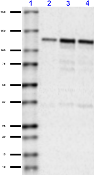
Western blot of purified anti-CD324 (E-Cadherin) antibody (clone 4A2). Lane 1: Molecular weight marker; Lane 2: 30 µg of human skin lysate; Lane 3: 30 µg of rat colon lysate. The blot was incubated with 5 µg/mL of the primary antibody overnight at 4°C, followed by incubation with HRP-labeled goat anti-mouse IgG (Cat. No. 405306). Enhanced chemiluminescence was used as the detection system. The top arrow indicates the full length E-Cadherin protein. The bottom arrow indicates the 33-kDa C-terminal fragment (CTF2). -

SeqIF™ (sequential immunofluorescence) staining on COMET™ of Purified anti-CD324 (E-Cadherin) (clone 4A2, yellow) on formalin-fixed paraffin-embedded human prostate carcinoma tissue at 5 µg/mL. Alexa Fluor™ Plus 647 Goat anti-Mouse IgG antibody (Lunaphore, Cat. No. DR647MS) was used as a secondary antibody. Nuclei were counterstained with DAPI (blue). Tissue underwent an all-in-one dewaxing and antigen retrieval preprocessing. -

IHC staining of Purified anti-CD324 (E-Cadherin) (clone 4A2) on formalin-fixed paraffin-embedded human tonsil tissue. Following antigen retrieval using Sodium Citrate H.I.E.R., 1X (Cat. No. 928502), the tissue was incubated without (panel A) and with (panel B) 10 µg/mL of Purified anti-CD324 (E-Cadherin) (clone 4A2) followed by incubation with Alexa Fluor® 647 Goat anti-mouse IgG (Cat. No. 405322) for 1 hour at room temperature. Nuclei were counterstained with DAPI (Cat. No. 422801). Images were captured with a 40X objective. Scale bar: 50 µm
| Cat # | Size | Price | Quantity Check Availability | Save | ||
|---|---|---|---|---|---|---|
| 866701 | 25 µg | 85€ | ||||
| 866702 | 100 µg | 221€ | ||||
E-cadherin is a calcium-dependent cell adhesion molecule (CAM) critical for formation and functions of adherent junctions during development and tumorigenesis. E-cadherin is usually expressed and localized at the surface of epithelial cells. However, it is also expressed in the adult and embryonic forebrain germinal zones in vivo and in cells derived from these regions growing in vitro. Conditional loss of E-cadherin in mouse brain causes defects in the self-renewal of neural stem cells both in vivo and in vitro. Overexpression of E-cadherin increases the number of neural stem cell colonies indicating that E-cadherin is required for the neural stem cell self-renewal function. In human glioblastoma tissue, the expression of E-cadherin is increased and usually correlates with unfavorable outcome.
Product DetailsProduct Details
- Verified Reactivity
- Human, Rat
- Antibody Type
- Monoclonal
- Host Species
- Mouse
- Formulation
- Phosphate-buffered solution, pH 7.2, containing 0.09% sodium azide.
- Preparation
- The antibody was purified by affinity chromatography.
- Concentration
- 0.5 mg/mL
- Storage & Handling
- The antibody solution should be stored undiluted between 2°C and 8°C.
- Application
-
WB - Quality tested
IHC-P - Verified
SB - Community verified - Recommended Usage
-
Each lot of this antibody is quality control tested by Western blotting. For Western blotting, the suggested use of this reagent is 1.0 - 10 µg per mL. For immunohistochemistry on formalin-fixed paraffin-embedded tissue sections, a concentration range of 1 - 10 µg/mL is suggested. It is recommended that the reagent be titrated for optimal performance for each application.
It is also recommended using EDTA-based solutions for dissociating attachment-dependent cell lines. - Application Notes
-
Lower MW bands detected in WB are cleaved C terminal fragments.
Cleavage of E-cadherin usually occurs in tumor development and cleaved E-cadherin fragments of ~29-38 kD often possess distinct oncogenic properties. - Additional Product Notes
-
For the use of this antibody in spatial biology applications, we have partnered with Lunaphore Technologies for demonstration of our antibodies on the COMET™. The COMET™ platform is an automated, end-to-end spatial biology solution developed for rapid and flexible multiplex tissue profiling. More information on the COMET™ and a complete list of our antibodies that have been demonstrated on the COMET™ can be found here.
- Application References
-
- David, J.M. and Rajasekaran, A.K. 2012. Cancer Res. 72(12): 2917–2923.
- Product Citations
-
- RRID
-
AB_2814610 (BioLegend Cat. No. 866701)
AB_2814611 (BioLegend Cat. No. 866702)
Antigen Details
- Structure
- Human CD324 (E-cadherin) is a 882 amino acid protein with a predicted molecular mass of 97 kD and observed molecular mass of ~120 kD.
- Distribution
-
Tissue distribution: Non-neural epithelial tissues and forebrain germinal zones
Cellular distribution: Golgi, plasma membrane, endosome, and cell - Function
- CD324 (E-cadherin) is involved in cell adhesion.
- Interaction
- Interacts with catenin family members
- Cell Type
- Embryonic Stem Cells, Epithelial cells, Neural Stem Cells
- Biology Area
- Cell Adhesion, Cell Biology, Immuno-Oncology, Immunology, Neuroscience, Stem Cells
- Molecular Family
- Adhesion Molecules, CD Molecules
- Antigen References
-
- Lewis-Tuffin LJ, et al. 2010. PLoS One. 5(10):e13665.
- Karpowicz P, et al. 2009. J Neurosci. 29(12):3885-96.
- Gene ID
- 999 View all products for this Gene ID
- UniProt
- View information about CD324 on UniProt.org

Related FAQs
- If an antibody clone has been previously successfully used in IBEX in one fluorescent format, will other antibody formats work as well?
-
It’s likely that other fluorophore conjugates to the same antibody clone will also be compatible with IBEX using the same sample fixation procedure. Ultimately a directly conjugated antibody’s utility in fluorescent imaging and IBEX may be specific to the sample and microscope being used in the experiment. Some antibody clone conjugates may perform better than others due to performance differences in non-specific binding, fluorophore brightness, and other biochemical properties unique to that conjugate.
- Will antibodies my lab is already using for fluorescent or chromogenic IHC work in IBEX?
-
Fundamentally, IBEX as a technique that works much in the same way as single antibody panels or single marker IF/IHC. If you’re already successfully using an antibody clone on a sample of interest, it is likely that clone will have utility in IBEX. It is expected some optimization and testing of different antibody fluorophore conjugates will be required to find a suitable format; however, legacy microscopy techniques like chromogenic IHC on fixed or frozen tissue is an excellent place to start looking for useful antibodies.
- Are other fluorophores compatible with IBEX?
-
Over 18 fluorescent formats have been screened for use in IBEX, however, it is likely that other fluorophores are able to be rapidly bleached in IBEX. If a fluorophore format is already suitable for your imaging platform it can be tested for compatibility in IBEX.
- The same antibody works in one tissue type but not another. What is happening?
-
Differences in tissue properties may impact both the ability of an antibody to bind its target specifically and impact the ability of a specific fluorophore conjugate to overcome the background fluorescent signal in a given tissue. Secondary stains, as well as testing multiple fluorescent conjugates of the same clone, may help to troubleshoot challenging targets or tissues. Using a reference control tissue may also give confidence in the specificity of your staining.
- How can I be sure the staining I’m seeing in my tissue is real?
-
In general, best practices for validating an antibody in traditional chromogenic or fluorescent IHC are applicable to IBEX. Please reference the Nature Methods review on antibody based multiplexed imaging for resources on validating antibodies for IBEX.
Other Formats
View All CD324 Reagents Request Custom Conjugation| Description | Clone | Applications |
|---|---|---|
| Purified anti-CD324 (E-Cadherin) | 4A2 | WB,IHC-P,SB |
| TotalSeq™-Bn1360 anti-CD324 (E-Cadherin) | 4A2 | SB |
Customers Also Purchased
Compare Data Across All Formats
This data display is provided for general comparisons between formats.
Your actual data may vary due to variations in samples, target cells, instruments and their settings, staining conditions, and other factors.
If you need assistance with selecting the best format contact our expert technical support team.
-
Purified anti-CD324 (E-Cadherin)

Western blot of purified anti-CD324 (E-Cadherin) antibody (c... 
SeqIF™ (sequential immunofluorescence) staining on COMET™ of... 
IHC staining of Purified anti-CD324 (E-Cadherin) (clone 4A2)... -
TotalSeq™-Bn1360 anti-CD324 (E-Cadherin)


Follow Us