 Login / Register


- Clone
- 143F (See other available formats)
- Regulatory Status
- RUO
- Other Names
- X-box binding protein 1
- Isotype
- Mouse IgG2a, κ
- Ave. Rating
- Submit a Review
- Product Citations
- publications

-

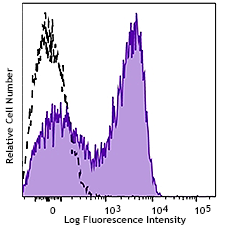
Human hepatocellular carcinoma cell line, HEPG2, was incubated overnight with (top) or without (bottom) thapsigargin (300 nM). Cells were then treated with FOXP3 Fix/Perm Buffer Set, and then stained with anti-Human Xbp-1s (clone 143F) PE (filled histogram) or mouse IgG2a, κ PE isotype control (open histogram). -

| Cat # | Size | Price | Quantity Check Availability | Save | ||
|---|---|---|---|---|---|---|
| 647503 | 25 tests | 123€ | ||||
| 647504 | 100 tests | 298€ | ||||
XBP-1 is a transcription factor containing a bZIP domain. It was first identified because of its ability to bind X-box, a conserved transcriptional element in the promoter of human HLA DR gene. XBP-1 has multiple functions. It controls MHC class II gene regulation and is also essential for differentiation of plasma cells. XBP-1 upregulates as part of the ER stress response, also known as the unfolded protein response. Unspliced XBP-1 is 261 amino acids and migrates on the SDS-PAGE around 33 kD; spliced XBP-1, recognized by clone 143F, is 371 amino acids and migrates around 55 kD.
Product DetailsProduct Details
- Verified Reactivity
- Human
- Antibody Type
- Monoclonal
- Host Species
- Mouse
- Immunogen
- XBP-1s recombinant protein
- Formulation
- Phosphate-buffered solution, pH 7.2, containing 0.09% sodium azide and BSA (origin USA)
- Preparation
- The antibody was purified by affinity chromatography and conjugated with PE under optimal conditions.
- Concentration
- Lot-specific (to obtain lot-specific concentration and expiration, please enter the lot number in our Certificate of Analysis online tool.)
- Storage & Handling
- The antibody solution should be stored undiluted between 2°C and 8°C, and protected from prolonged exposure to light. Do not freeze.
- Application
-
ICFC - Quality tested
- Recommended Usage
-
Each lot of this antibody is quality control tested by intracellular immunofluorescent staining with flow cytometric analysis. For flow cytometric staining, the suggested use of this reagent is 5 µl per million cells in 100 µl staining volume or 5 µl per 100 µl of whole blood.
- Excitation Laser
-
Blue Laser (488 nm)
Green Laser (532 nm)/Yellow-Green Laser (561 nm)
- Application Notes
-
Additional reported applications (for the relevant formats) include: immunofluorescence14 and immunohistochemical staining of PFA-fixed paraffin-embedded sections1,14.
- Application References
-
- Tsang KY, et al. 2007. PLOS Biol. 5:568. (IHC) PubMed
- Farhan H, et al. 2008. EMBO J. 27:2043 (WB) PubMed
- Farhan H, et al. 2010. J. Cell Biol. 189:997. PubMed
- Feng-Jin G, et al. 2010. Cell Signal. [Epub ahead of print]
- Wang L, et al. 2011. Hum Mol Genet. PubMed
- Li J, et al. 2011. J. Biol Chem. 286:4912. PubMed
- Wang L, et al. 2011. Hum Mol Genet. 20:1008. PubMed
- Liu Y, et al. 2011. J Biol Chem. 286:13161. PubMed
- Shirley CM, et al. 2011 Blood. 117:6297. PubMed
- Vidal RL., et al. 2012. Hum Mol Genet. PubMed
- Byrd AE, et al. 2012. J Cell Biol. 196:689. PubMed
- Dickie LJ, et al. 2012. Ann rheum Dis. 71:2035. PubMed
- Bonetti P, et al. 2013. Blood. 122:2233. PubMed
- Maestre L, et al. 2009. Haematologica. 94:419. (IF, IHC)
- Rodriguez M, et al. 2014. J Biol Chem. 289:22942. PubMed
- Product Citations
-
- RRID
-
AB_2563447 (BioLegend Cat. No. 647503)
AB_2563448 (BioLegend Cat. No. 647504)
Antigen Details
- Distribution
-
Ubiquitously expressed.
- Function
- Upregulated by ER stress, IL-6, and IL-4. Downregulation correlates with tumor progression in prostate cancer. Downregulated by PAX5 transcription factor.
- Biology Area
- Cell Biology
- Antigen References
-
1. Liou HC, et al. 1990. Science 247:1581.
2. Ponath PD, et al. 1993. J. Biol. Chem.268:17074.
3. Takahashi S, et al. 2002. Prostate 50:154. - Gene ID
- 7494 View all products for this Gene ID
- UniProt
- View information about XBP-1s on UniProt.org


Related FAQs
- What type of PE do you use in your conjugates?
- We use R-PE in our conjugates.
Other Formats
View All XBP-1s Reagents Request Custom Conjugation| Description | Clone | Applications |
|---|---|---|
| Purified anti-XBP-1s | 143F | WB,ChIP,ICC,IHC-P |
| PE anti-XBP-1s | 143F | ICFC |
| Alexa Fluor® 647 anti-XBP-1s | 143F | ICFC |
Customers Also Purchased

Compare Data Across All Formats
This data display is provided for general comparisons between formats.
Your actual data may vary due to variations in samples, target cells, instruments and their settings, staining conditions, and other factors.
If you need assistance with selecting the best format contact our expert technical support team.
-
Purified anti-XBP-1s

Extracts of untreated HepG2 cells (lane 1) and extracts of H... 
Chromatin Immunoprecipitations (ChIP) were performed using f... 
Total cell lysates (15 µg protein) from HepG2 cells treated ... -
PE anti-XBP-1s

Human hepatocellular carcinoma cell line, HEPG2, was incubat... 
-
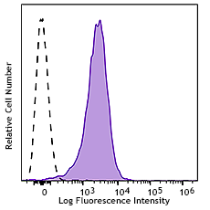
Alexa Fluor® 647 anti-XBP-1s

Human hepatocellular carcinoma cell line, HEPG2, was incubat... 


Follow Us