 Login / Register


- Clone
- LG.3A10 (See other available formats)
- Regulatory Status
- RUO
- Other Names
- T14, S152, Tp55, TNFRSF7
- Isotype
- Armenian Hamster IgG
- Ave. Rating
- Submit a Review
- Product Citations
- publications

-

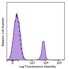
C57BL/6 mouse splenocytes stained with LG.3A10 PE
| Cat # | Size | Price | Quantity Check Availability | Save | ||
|---|---|---|---|---|---|---|
| 124209 | 50 µg | 94€ | ||||
| 124210 | 200 µg | 268€ | ||||
CD27 is also known as S152 and T14. A member of the tumor necrosis factor receptor (TNFR) superfamily, it is a 45 kD protein expressed on peripheral T cells, memory B cells, NK cells, and thymocyte subset. Through its ligand, CD70, CD27 plays a key role in T cell and B cell interactions. Additionally, ligation of CD27 on naïve T cells may be important in their maturation to effector cells.
Product DetailsProduct Details
- Verified Reactivity
- Mouse, Rat, Human
- Antibody Type
- Monoclonal
- Host Species
- Armenian Hamster
- Immunogen
- Armenian hamster fibroblast line ARHO12 transfected with mouse CD27 cDNA
- Formulation
- Phosphate-buffered solution, pH 7.2, containing 0.09% sodium azide
- Preparation
- The antibody was purified by affinity chromatography, and conjugated with PE under optimal conditions.
- Concentration
- 0.2 mg/ml
- Storage & Handling
- The antibody solution should be stored undiluted between 2°C and 8°C, and protected from prolonged exposure to light. Do not freeze.
- Application
-
FC - Quality tested
- Recommended Usage
-
Each lot of this antibody is quality control tested by immunofluorescent staining with flow cytometric analysis. For flow cytometric staining, the suggested use of this reagent is ≤0.25 µg per million cells in 100 µl volume. It is recommended that the reagent be titrated for optimal performance for each application.
- Excitation Laser
-
Blue Laser (488 nm)
Green Laser (532 nm)/Yellow-Green Laser (561 nm)
- Application Notes
-
Additional reported applications (for the relevant formats) include: functional assay1,3 and immunohistomecial staining of acetone-fixed frozen sections2. The Ultra-LEAF™ purified antibody (Endotoxin < 0.01 EU/µg, Azide-Free, 0.2 µm filtered) is recommended for functional assays (Cat. Nos. 124243 & 124244).
- Application References
-
- Gravestein LA, et al. 1995. Int. Immunol. 7:551. (FA)
- Gravestein LA, et al. 1996. J. Exp. Med. 184:675. (IHC)
- Takeda K, et al. 2000. J. Immunol. 164:1741. (FA)
- Welner RS, et al. 2009. J. Immunol. 183:7768. PubMed
- Vicetti Miguel RC, et al. 2012. J. Immunol. 189:3449. PubMed
- White CA, et al. 2014. J Immunol. 193:5933. PubMed
- Iwata S, et al. 2015. Lupus. 24:695. PubMed
- Product Citations
-
- RRID
-
AB_1236464 (BioLegend Cat. No. 124209)
AB_1236459 (BioLegend Cat. No. 124210)
Antigen Details
- Structure
- TNFR superfamily, 45 kD
- Distribution
-
Peripheral blood T cells, memory B cells, NK cells, thymocyte subset
- Function
- Costimulatory signal for T and B cell activation, T cell development
- Interaction
- TRAF2, TRAF5, CD27-binding protein (Siva)
- Ligand/Receptor
- CD70
- Cell Type
- B cells, NK cells, T cells, Thymocytes
- Biology Area
- Costimulatory Molecules, Immunology
- Molecular Family
- CD Molecules
- Antigen References
-
1. Hintzen RQ, et al. 1994. Immunol. Today 15:307.
2. Akiba H, et al. 1998. J. Biol. Chem. 273:13353.
3. Kobata T, et al. 1995. P. Natl. Acad. Sci. USA 92:11249.
4. Hendriks J, et al. 2000. Nat. Immunol. 1:433
5. Hintzen RQ, et al. 1994. Int. Immunol. 6:477
6. Camerini D, et al. 1991. J. Immunol. 147:3165 - Gene ID
- 21940 View all products for this Gene ID 939 View all products for this Gene ID 500318 View all products for this Gene ID
- UniProt
- View information about CD27 on UniProt.org
Related Pages & Pathways
Pathways


Related FAQs
- What type of PE do you use in your conjugates?
- We use R-PE in our conjugates.
Other Formats
View All CD27 Reagents Request Custom ConjugationCustomers Also Purchased
Compare Data Across All Formats
This data display is provided for general comparisons between formats.
Your actual data may vary due to variations in samples, target cells, instruments and their settings, staining conditions, and other factors.
If you need assistance with selecting the best format contact our expert technical support team.
-
Purified anti-mouse/rat/human CD27

C57BL/6 mouse splenocytes stained with purified LG.3A10, fol... -
Biotin anti-mouse/rat/human CD27

C57BL/6 mouse splenocytes stained with biotinylated LG.3A10,... -
FITC anti-mouse/rat/human CD27

C57BL/6 mouse splenocytes stained with LG.3A10 FITC -
PE anti-mouse/rat/human CD27

C57BL/6 mouse splenocytes stained with LG.3A10 PE -
APC anti-mouse/rat/human CD27

C57BL/6 mouse splenocytes stained with LG.3A10 APC -
PerCP/Cyanine5.5 anti-mouse/rat/human CD27

C57BL/6 mouse splenocytes were stained with CD45R/B220 APC a... -
PE/Cyanine7 anti-mouse/rat/human CD27

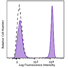
C57BL/6 mouse thymocytes stained with LG.3A10 PE/Cyanine7 -
Pacific Blue™ anti-mouse/rat/human CD27

C57Bl/6 mouse splenocytes were stained with CD27 (clone LG.3... -
Alexa Fluor® 647 anti-mouse/rat/human CD27

C57Bl/6 mouse splenocytes were stained with CD27 (clone LG.3... -
Alexa Fluor® 488 anti-mouse/rat/human CD27

C57Bl/6 mouse splenocytes were stained with CD27 (clone LG.3... -
Brilliant Violet 421™ anti-mouse/rat/human CD27

C57Bl/6 mouse splenocytes were stained with CD27 (clone LG.3... -
APC/Cyanine7 anti-mouse/rat/human CD27

C57BL/6 mouse splenocytes were stained with CD45R/B220 APC a... -
PE/Dazzle™ 594 anti-mouse/rat/human CD27

C57B/6 mouse splenocytes were stained with CD27 (clone LG.3A... -
Brilliant Violet 510™ anti-mouse/rat/human CD27

C57B/6 mouse splenocytes were stained with CD27 (clone LG.3A... -
Brilliant Violet 650™ anti-mouse/rat/human CD27


-
TotalSeq™-A0191 anti-mouse/rat/human CD27
-
Brilliant Violet 785™ anti-mouse/rat/human CD27

C57BL/6 mouse splenocytes were stained with CD45R/B220 PE an... -
APC/Fire™ 750 anti-mouse/rat/human CD27

C57BL/6 mouse splenocytes were stained with CD45R/B220 PE an... -
Alexa Fluor® 700 anti-mouse/rat/human CD27

C57BL/6 mouse splenocytes were stained with CD45R/B220 PE an... -
Ultra-LEAF™ Purified anti-mouse/rat/human CD27

C57BL/6 mouse splenocytes stained with purified LG.3A10, fol... -
TotalSeq™-C0191 anti-mouse/rat/human CD27
-
TotalSeq™-B0191 anti-mouse/rat/human CD27
-
Brilliant Violet 605™ anti-mouse/rat/human CD27

C57BL/6 mouse splenocytes were stained with CD45R/B220 APC a...


Follow Us