 Login / Register


- Clone
- MEC13.3 (See other available formats)
- Regulatory Status
- RUO
- Other Names
- PECAM-1, EndoCAM
- Isotype
- Rat IgG2a, κ
- Ave. Rating
- Submit a Review
- Product Citations
- publications

-

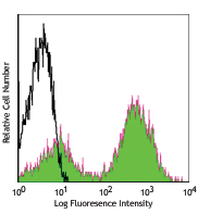
C57BL/6 mouse splenocytes stained with MEC13. 3 PE
| Cat # | Size | Price | Quantity Check Availability | Save | ||
|---|---|---|---|---|---|---|
| 102507 | 50 µg | 98€ | ||||
| 102508 | 200 µg | 235€ | ||||
CD31 is a 130-140 kD glycoprotein, also known as platelet endothelial cell adhesion molecule (PECAM-1), EndoCAM, and gpIIa. It is a member of the Ig superfamily, expressed on endothelial cells, platelets, granulocytes, monocytes/macrophages, dendritic cells, and T and B cell subsets, and is critical for cell-to-cell interactions. The primary ligands for CD31 have been reported to be CD38 and the vitronectin receptor (αv β3 integrin, CD51/CD61). Other reported functions of CD31 are neutrophil emigration to sites of inflammation, and angiogenesis.
Product DetailsProduct Details
- Verified Reactivity
- Mouse
- Antibody Type
- Monoclonal
- Host Species
- Rat
- Immunogen
- Polyoma middle T transformed EC line tEnd.1
- Formulation
- Phosphate-buffered solution, pH 7.2, containing 0.09% sodium azide.
- Preparation
- The antibody was purified by affinity chromatography, and conjugated with PE under optimal conditions.
- Concentration
- 0.2 mg/ml
- Storage & Handling
- The antibody solution should be stored undiluted between 2°C and 8°C, and protected from prolonged exposure to light. Do not freeze.
- Application
-
FC - Quality tested
- Recommended Usage
-
Each lot of this antibody is quality control tested by immunofluorescent staining with flow cytometric analysis. For flow cytometric staining, the suggested use of this reagent is ≤1.0 µg per million cells in 100 µl volume. It is recommended that the reagent be titrated for optimal performance for each application.
- Excitation Laser
-
Blue Laser (488 nm)
Green Laser (532 nm)/Yellow-Green Laser (561 nm)
- Application Notes
-
Anti-mouse CD31 clones 390 and MEC13.3 bind to their respective non-overlapping epitopes in IgD2 of CD31.8 Additional reported applications (in the relevant formats) include: immunoprecipitation1, in vitro and in vivo blocking of CD31-mediated cell-cell interactions1-4, immunohistochemical staining1,5,6 of acetone-fixed frozen sections and zinc-fixed paraffin-embedded sections, and spatial biology (IBEX)12,13.
Special Note: The antibody works well on acetone-fixed frozen sections as well as Zinc-fixed paraffin-embedded sections. It sometime works on formalin-fixed and paraformaldehyde-fixed paraffin-embedded tissue sections but inconsistent results have been reported. This antibody is not recommended for formalin-fixed paraffin-embedded sections or for Western blot analysis. The Ultra-LEAF™ Purified antibody (Endotoxin < 0.01 EU/µg, Azide-Free, 0.2 µm filtered) is recommended for functional assays (Cat. No. 102529 and 102530). - Application References
-
- Vecchi A, et al. 1994. Eur. J. Cell Biol. 63:247. (IP, IHC, Block)
- Christofidou-Solomidou M, et al. 1997. J. Immunol. 158:4872. (Block)
- DeLisser HM, et al. 1997. Am. J. Pathol. 151:671. (Block)
- Rosenblum WI, et al. 1994. Am. J. Pathol. 145:33. (Block)
- Baldwin HS, et al. 1994. Development 120:2539. (IHC)
- Voswinckel R, et al. 2003. Circ. Res. 93:372. (IHC)
- Leung VW, et al. 2009. Am J. Pathol. 175:1757. PubMed
- Chacko AM, et al. 2012. PLoS One 7:e34958.
- Giacomini C, et al. 2014. Exp Eye Res. 18:1. PubMed
- Morita R, et al. 2015. PNAS. 112:160. PubMed
- Ito A, et al. 2015. Brain Res. 1594:310. PubMed
- Radtke AJ, et al. 2020. Proc Natl Acad Sci U S A. 117:33455-65. (SB) PubMed
- Radtke AJ, et al. 2022. Nat Protoc. 17:378-401. (SB) PubMed
- Product Citations
-
- RRID
-
AB_312914 (BioLegend Cat. No. 102507)
AB_312915 (BioLegend Cat. No. 102508)
Antigen Details
- Structure
- Ig superfamily, 130-140 kD
- Distribution
-
Endothelial cells, platelets, granulocytes, monocytes/macrophages, dendritic cells, T and B cell subsets
- Function
- Adhesion
- Ligand/Receptor
- CD38, αV/β3 integrin
- Cell Type
- B cells, Dendritic cells, Endothelial cells, Granulocytes, Macrophages, Monocytes, Neutrophils, Platelets, T cells
- Biology Area
- Angiogenesis, Cell Adhesion, Cell Biology, Immunology, Neuroinflammation, Neuroscience
- Molecular Family
- Adhesion Molecules, CD Molecules
- Antigen References
-
1. Barclay AN, et al. 1997. The Leukocyte Antigen FactsBook Academic Press.
2. DeLisser HM, et al. 1994. Immunol. Today 15:490.
3. Newman PJ, et al. 1990. Science 247:1219. - Gene ID
- 18613 View all products for this Gene ID
- UniProt
- View information about CD31 on UniProt.org
Related Pages & Pathways
Pathways


Related FAQs
- What type of PE do you use in your conjugates?
- We use R-PE in our conjugates.
Other Formats
View All CD31 Reagents Request Custom Conjugation| Description | Clone | Applications |
|---|---|---|
| APC anti-mouse CD31 | MEC13.3 | FC |
| Biotin anti-mouse CD31 | MEC13.3 | FC |
| FITC anti-mouse CD31 | MEC13.3 | FC |
| PE anti-mouse CD31 | MEC13.3 | FC |
| Purified anti-mouse CD31 | MEC13.3 | FC,IP,Block,IHC-F |
| Alexa Fluor® 488 anti-mouse CD31 | MEC13.3 | FC |
| Alexa Fluor® 647 anti-mouse CD31 | MEC13.3 | FC,3D IHC,SB |
| Alexa Fluor® 594 anti-mouse CD31 | MEC13.3 | FC,IHC-F,3D IHC,SB |
| PerCP/Cyanine5.5 anti-mouse CD31 | MEC13.3 | FC |
| PE/Cyanine7 anti-mouse CD31 | MEC13.3 | FC |
| PE/Dazzle™ 594 anti-mouse CD31 | MEC13.3 | FC |
| APC/Fire™ 750 anti-mouse CD31 | MEC13.3 | FC |
| Ultra-LEAF™ Purified anti-mouse CD31 | MEC13.3 | FC,IP,Block,IHC-F |
| APC/Cyanine7 anti-mouse CD31 | MEC13.3 | FC |
| Spark YG™ 570 anti-mouse CD31 | MEC13.3 | IHC-F |
| Spark Red™ 718 anti-mouse CD31 (Flexi-Fluor™) | MEC13.3 | FC |
Customers Also Purchased
Compare Data Across All Formats
This data display is provided for general comparisons between formats.
Your actual data may vary due to variations in samples, target cells, instruments and their settings, staining conditions, and other factors.
If you need assistance with selecting the best format contact our expert technical support team.
-
APC anti-mouse CD31

C57BL/6 splenocytes stained with MEC13.3 APC -
Biotin anti-mouse CD31

C57BL/6 mouse splenocytes stained with biotinylated MEC13.3,... -
FITC anti-mouse CD31

C57BL/6 mouse splenocytes stained with MEC13.3 FITC -
PE anti-mouse CD31

C57BL/6 mouse splenocytes stained with MEC13. 3 PE -
Purified anti-mouse CD31

C57BL/6 mouse splenocytes stained with purified MEC13.3, fol... -
Alexa Fluor® 488 anti-mouse CD31

C57BL/6 mouse splenocytes stained with MEC13.3 Alexa Fluor® ... -
Alexa Fluor® 647 anti-mouse CD31

C57BL/6 mouse splenocytes stained with MEC13.3 Alexa Fluor® ... 
Formalin-fixed, 300 micron-thick mouse kidney section was bl... 
In-vivo mouse testis labeling of blood vessels with Alexa Fl... 
Paraformaldehyde-fixed (4%), 500 μm-thick mouse kidney secti... 
Confocal image of C57BL/6 mouse small intestine sample acqui... 
Confocal image of C57BL/6 mouse small intestine sample acqui... -
Alexa Fluor® 594 anti-mouse CD31

C57BL/6 mouse splenocytes were stained with CD31 (clone MEC1... 
C57BL/6 mouse frozen liver section was fixed with 4% parafor... 
Dissected C57/B6 mouse stomach was immersed in 4% paraforma... 
Paraformaldehyde-fixed (4%), 500 μm-thick mouse kidney secti... 
Confocal image of C57BL/6 mouse lung sample acquired using t... 
Confocal image of C57BL/6 mouse lung sample acquired using t... -
PerCP/Cyanine5.5 anti-mouse CD31

C57BL/6 mouse splenocytes were stained with CD31 (clone MEC1... -
PE/Cyanine7 anti-mouse CD31

C57BL/6 mouse splenocytes were stained with CD31 (clone MEC1... -
PE/Dazzle™ 594 anti-mouse CD31

MEC13dot3_PEDZZL594_CD31_Antibody_040517 -
APC/Fire™ 750 anti-mouse CD31

C57BL/6 were stained with CD31 (clone MEC13.3) APC/Fire™ 750... -
Ultra-LEAF™ Purified anti-mouse CD31
-
APC/Cyanine7 anti-mouse CD31

C57BL/6 mouse splenocytes were stained with CD31 (clone MEC1... -
Spark YG™ 570 anti-mouse CD31

C57BL/6 mouse frozen lymph node section was fixed with 4% pa... -
Spark Red™ 718 anti-mouse CD31 (Flexi-Fluor™)


Follow Us