 Login / Register


- Clone
- 5B11 (See other available formats)
- Regulatory Status
- RUO
- Other Names
- IL-3 Receptor α chain, IL-3Rα
- Isotype
- Rat IgG2a, κ
- Ave. Rating
- Submit a Review
- Product Citations
- publications

-

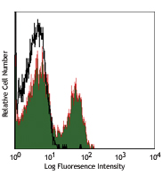
C57BL/6 mouse bone marrow cells stained with 5B11 PE and CD11b FITC
| Cat # | Size | Price | Quantity Check Availability | Save | ||
|---|---|---|---|---|---|---|
| 106005 | 25 µg | 69€ | ||||
CD123 is a 70 kD α chain subunit of the IL-3 receptor (IL-3R α). It is a member of the immunoglobulin superfamily that is expressed on hematopoietic progenitors, basophils, mast cells, and megakaryocytes. This transmembrane glycoprotein can bind IL-3 with low affinity but cannot transduce signals without association with additional protein partners. CD123 can complex with either the common β chain (CDw131) or the IL-3R β chain (AIC2A) to form high-affinity heterodimeric IL-3 receptors. CDw131 can complex with the α subunits of the mouse IL-3R, IL-5R and GM-CSFR to form high-affinity receptors, while the IL-3 R β subunit is specific for IL-3 but binds with low affinity. IL-3 binding to the receptor complex can induce proliferation and differentiation of hematopoietic cells. The 5B11 antibody does not block binding of IL-3 to the high affinity IL-3 receptor.
Product DetailsProduct Details
- Verified Reactivity
- Mouse
- Antibody Type
- Monoclonal
- Host Species
- Rat
- Immunogen
- CTLL cells transfected with the mouse IL-3Rα chain
- Formulation
- Phosphate-buffered solution, pH 7.2, containing 0.09% sodium azide.
- Preparation
- The antibody was purified by affinity chromatography, and conjugated with PE under optimal conditions.
- Concentration
- 0.2 mg/ml
- Storage & Handling
- The antibody solution should be stored undiluted between 2°C and 8°C, and protected from prolonged exposure to light. Do not freeze.
- Application
-
FC - Quality tested
- Recommended Usage
-
Each lot of this antibody is quality control tested by immunofluorescent staining with flow cytometric analysis. For flow cytometric staining, the suggested use of this reagent is ≤1.0 µg per million cells in 100 µl volume. It is recommended that the reagent be titrated for optimal performance for each application.
- Excitation Laser
-
Blue Laser (488 nm)
Green Laser (532 nm)/Yellow-Green Laser (561 nm)
- Application Notes
-
For most successful immunofluorescent staining results, it may be important to maximize signal over background by using a relatively bright fluorochrome-antibody conjugate (Cat. No. 106006) or by using a high sensitivity, three-layer staining technique (e.g., including a biotinylated antibody (Cat. No. 106004) or biotinylated anti-rat IgG second step (Cat. No. 405402), followed by SAv-PE (Cat. No. 405204)). To reduce non-specific binding to cells bearing Fc-receptors, pre-incubation of cells with anti-mouse CD16/CD32, clone 93 (Cat. No. 101301/101302), is recommended prior to immunofluorescent staining.
- Application References
-
- Ichihara, et al. 1995. EMBO J. 14:939. (FC)
- Mueller DL, et al. 1994. J. Immunol. 153:3014. (FC)
- Cervantes-Barragan L, et al. 2007. Blood 109:1131. (FC)
- Product Citations
-
- RRID
-
AB_2124403 (BioLegend Cat. No. 106005)
Antigen Details
- Structure
- Ig superfamily, associates with CDw131 (common β chain) or IL-3 receptor β chain, 70 kD
- Distribution
-
Hematopoietic progenitors, basophils, mast cells, megakaryocytes
- Function
- Proliferation, differentiation of hematopoietic cells
- Ligand/Receptor
- IL-3
- Cell Type
- Basophils, Dendritic cells, Hematopoietic stem and progenitors, Mast cells, Megakaryocytes
- Biology Area
- Immunology
- Molecular Family
- CD Molecules, Cytokine/Chemokine Receptors
- Antigen References
-
1. Barclay A, et al. 1997. The Leukocyte Antigen FactsBook Academic Press.
2. Miyajima A, et al. 1993. Blood 82:1960.
3. Itoh N, et al. 1990. Science 247:324. - Gene ID
- 16188 View all products for this Gene ID
- UniProt
- View information about CD123 on UniProt.org
Related Pages & Pathways
Pathways


Related FAQs
- What type of PE do you use in your conjugates?
- We use R-PE in our conjugates.
Other Formats
View All CD123 Reagents Request Custom Conjugation| Description | Clone | Applications |
|---|---|---|
| Biotin anti-mouse CD123 | 5B11 | FC |
| PE anti-mouse CD123 | 5B11 | FC |
| Purified anti-mouse CD123 | 5B11 | FC |
Customers Also Purchased

Compare Data Across All Formats
This data display is provided for general comparisons between formats.
Your actual data may vary due to variations in samples, target cells, instruments and their settings, staining conditions, and other factors.
If you need assistance with selecting the best format contact our expert technical support team.
-
Biotin anti-mouse CD123

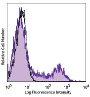
C57BL/6 bone marrow cells double stained with CD11b FITC and... 
C57BL/6 bone marrow cells double stained with rat IgG2a (Clo... -
PE anti-mouse CD123

C57BL/6 mouse bone marrow cells stained with 5B11 PE and CD1... -
Purified anti-mouse CD123
C57BL/6 mouse bone marrow cells stained with 5B11 PE and CD1...


Follow Us