 Login / Register


- Clone
- BJ18 (See other available formats)
- Regulatory Status
- RUO
- Workshop
- VI A034
- Other Names
- Hermes, Pgp-1, H-CAM, HUTCH-1, ECMR III, gp85, Ly-24
- Isotype
- Mouse IgG1, κ
- Ave. Rating
- Submit a Review
- Product Citations
- publications

-

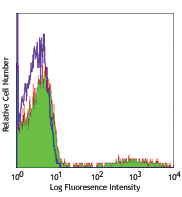
Human peripheral blood lymphocytes stained with BJ18 FITC
| Cat # | Size | Price | Quantity Check Availability | Save | ||
|---|---|---|---|---|---|---|
| 338803 | 25 tests | 67€ | ||||
| 338804 | 100 tests | 151€ | ||||
CD44 is a 80-95 kD glycoprotein also known as Hermes, Pgp1, H-CAM, or HUTCH. It is expressed on all leukocytes, endothelial cells, hepatocytes, and mesenchymal cells. As B and T cells become activated or progress to the memory stage, CD44 expression increases from a low or mid level of intensity to high expression levels. Thus, CD44 has been reported to be a valuable marker for memory cell subsets. CD44 is an adhesion molecule involved in leukocyte attachment to and rolling on endothelial cells, homing to peripheral lymphoid organs and to the sites of inflammation, and leukocyte aggregation.
Product DetailsProduct Details
- Verified Reactivity
- Human
- Reported Reactivity
- African Green, Baboon, Cynomolgus, Rhesus
- Antibody Type
- Monoclonal
- Host Species
- Mouse
- Immunogen
- Normal human PBL
- Formulation
- Phosphate-buffered solution, pH 7.2, containing 0.09% sodium azide and BSA (origin USA)
- Preparation
- The antibody was purified by affinity chromatography and conjugated with FITC under optimal conditions.
- Concentration
- Lot-specific (to obtain lot-specific concentration and expiration, please enter the lot number in our Certificate of Analysis online tool.)
- Storage & Handling
- The antibody solution should be stored undiluted between 2°C and 8°C, and protected from prolonged exposure to light. Do not freeze.
- Application
-
FC - Quality tested
- Recommended Usage
-
Each lot of this antibody is quality control tested by immunofluorescent staining with flow cytometric analysis. For flow cytometric staining, the suggested use of this reagent is 5 µl per million cells in 100 µl staining volume or 5 µl per 100 µl of whole blood.
- Excitation Laser
-
Blue Laser (488 nm)
- Application References
-
- Kishimoto T, et al. eds. 1997 Leucocyte Typing VI:White Cell Differentiation Antigen. Garland Publishing Inc.
- Product Citations
-
- RRID
-
AB_1501204 (BioLegend Cat. No. 338803)
AB_1501197 (BioLegend Cat. No. 338804)
Antigen Details
- Structure
- Variable splicing of CD44 gene generates many CD44 isoforms, 85 kD
- Distribution
-
All leukocytes, epithelial cells, endothelial cells, hepatocytes, mesenchymal cells
- Function
- Leukocyte attachment and rolling on endothelial cells, stromal cells and ECM
- Ligand/Receptor
- Hyaluronan, MIP-1β, fibronectin, collagen
- Cell Type
- Endothelial cells, Epithelial cells, Leukocytes, Mesenchymal cells, Mesenchymal Stem Cells, Tregs
- Biology Area
- Cell Adhesion, Cell Biology, Immunology, Stem Cells
- Molecular Family
- Adhesion Molecules, CD Molecules
- Antigen References
-
1. Barclay AN, et al. 1997. The Leukocyte Antigen FactsBook Academic Press.
2. Haynes BF, et al. 1991. Cancer Cells 3:347.
3. Goldstein LA, et al. 1989. Cell 56:1063.
4. Mikecz K, et al. 1995. Nat. Med. 1:558. - Gene ID
- 960 View all products for this Gene ID
- UniProt
- View information about CD44 on UniProt.org
Related Pages & Pathways
Pathways


Other Formats
View All CD44 Reagents Request Custom Conjugation| Description | Clone | Applications |
|---|---|---|
| Purified anti-human CD44 | BJ18 | FC,CyTOF®,ICC |
| FITC anti-human CD44 | BJ18 | FC |
| APC anti-human CD44 | BJ18 | FC |
| PE anti-human CD44 | BJ18 | FC |
| Brilliant Violet 421™ anti-human CD44 | BJ18 | FC |
| Purified anti-human CD44 (Maxpar® Ready) | BJ18 | FC,CyTOF® |
| Alexa Fluor® 700 anti-human CD44 | BJ18 | FC |
| PE/Dazzle™ 594 anti-human CD44 | BJ18 | FC |
| PE/Cyanine7 anti-human CD44 | BJ18 | FC |
| APC/Fire™ 750 anti-human CD44 | BJ18 | FC |
| PerCP/Cyanine5.5 anti-human CD44 | BJ18 | FC |
| Pacific Blue™ anti-human CD44 | BJ18 | FC |
| TotalSeq™-A0125 anti-human CD44 | BJ18 | PG |
| TotalSeq™-C0125 anti-human CD44 | BJ18 | PG |
| Alexa Fluor® 488 anti-human CD44 | BJ18 | FC |
| Brilliant Violet 785™ anti-human CD44 | BJ18 | FC |
| TotalSeq™-B0125 anti-human CD44 Antibody | BJ18 | PG |
| TotalSeq™-D0125 anti-human CD44 | BJ18 | PG |
Customers Also Purchased
Compare Data Across All Formats
This data display is provided for general comparisons between formats.
Your actual data may vary due to variations in samples, target cells, instruments and their settings, staining conditions, and other factors.
If you need assistance with selecting the best format contact our expert technical support team.
-
Purified anti-human CD44

MDA-MB-231 cells were stained with anti-CD44 (clone BJ18), f... -
FITC anti-human CD44

Human peripheral blood lymphocytes stained with BJ18 FITC -
APC anti-human CD44

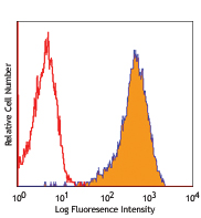
Human peripheral blood lymphocytes stained with BJ18 APC -
PE anti-human CD44

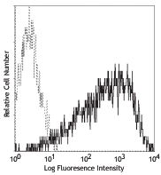
Human peripheral blood lymphocytes stained with BJ18 PE 
Pre-lysed human blood leukocytes were stained with CD44 (clo... -
Brilliant Violet 421™ anti-human CD44

Human peripheral blood lymphocytes were stained with CD44 (c... -
Purified anti-human CD44 (Maxpar® Ready)

Human PBMCs stained with 166Er-anti-CD44 (BJ18) and 158Gd-an... -
Alexa Fluor® 700 anti-human CD44

Human peripheral blood lymphocytes were stained with CD44 (c... -
PE/Dazzle™ 594 anti-human CD44

Human peripheral blood lymphocytes were stained with CD44 (c... -
PE/Cyanine7 anti-human CD44

Human peripheral blood lymphocytes were stained with CD44 (c... -
APC/Fire™ 750 anti-human CD44

Human peripheral blood lymphocytes were stained with CD44 (c... -
PerCP/Cyanine5.5 anti-human CD44

Human peripheral blood lymphocytes were stained with CD44 (c... -
Pacific Blue™ anti-human CD44

Human peripheral blood lymphocytes were stained with CD44 (c... -
TotalSeq™-A0125 anti-human CD44
-
TotalSeq™-C0125 anti-human CD44
-
Alexa Fluor® 488 anti-human CD44

Human peripheral blood lymphocytes were stained with CD3 APC... -
Brilliant Violet 785™ anti-human CD44

Human peripheral blood lymphocytes were stained with CD44 (c... -
TotalSeq™-B0125 anti-human CD44 Antibody
-
TotalSeq™-D0125 anti-human CD44


Follow Us