 Login / Register


- Regulatory Status
- RUO
- Other Names
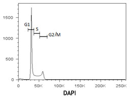
- live cell cycle
- Ave. Rating
- Submit a Review
- Product Citations
- publications

-

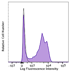
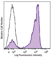
Ramos cells treated with 5µM CytoPhase™ Violet dye for 90 minutes at 37°C. Cells were then acquired on a flow cytometer equipped with a 405 nm laser with a 450/50 bandpass filter.
| Cat # | Size | Price | Quantity Check Availability | Save | ||
|---|---|---|---|---|---|---|
| 425701 | 200 µL | 226€ | ||||
CytoPhase™ Violet is a cell-permeant DNA-binding dye with the molecular weight of 560.96D and can be excited by either UV (355 nm or 375 nm) or violet laser (405 nm). Its emission maximum wavelength is about 440nm. It can be used for flow cytometric analysis of cell cycle in live or fixed cells. This dye can also be used for counterstaining the nuclei of cells in fluorescent microscopic imaging.
Product DetailsProduct Details
- Formulation
- 5mM solution in H20 (200 µl/vial)
- Concentration
- 5.0 mM
- Storage & Handling
- Upon receipt, CytoPhase™ Violet should be stored at 4°C and protected from light.
- Application
-
FC - Quality tested
- Recommended Usage
-
The recommended usage concentration for CytoPhase™ Violet is 5µM - 10µM.
- Application Notes
-
Prepare cells of interest in appropriate cell culture media at a concentration of 0.5-1x 10⁶ cells /ml. Add CytoPhase™ Violet dye to the cell suspension to bring it to 5µM (1:1000)-10µM (1:500) working solution and incubate for 90 minutes at 37°C (CO2 incubator). Optimal concentration might require titration of the dye based on differing cell types. Transfer cells to a FACS tube (cells do not require a wash step) and acquire samples on a flow cytometer equipped with a violet laser.
CytoPhase™ Violet can be used concurrently with antibody staining procedures.
Antigen Details
- Distribution
-
Nucleus, DNA.
- Biology Area
- Cell Biology, Cell Cycle/DNA Replication, Cell Proliferation and Viability
- Gene ID
- NA


Follow Us