 Login / Register


- Clone
- 29F.1A12 (See other available formats)
- Regulatory Status
- RUO
- Other Names
- PD-1, Programmed Death-1, PDCD1
- Isotype
- Rat IgG2a, κ
- Ave. Rating
- Submit a Review
- Product Citations
- publications

-

Con-A and IL-2 stimulated C57BL/6 splenocytes (3 days) were stained with CD3 (clone 145-2C11) FITC and CD279 (clone 29F.1A12) Brilliant Violet 785™ (top), or rat IgG2a, κ Brilliant Violet 785™ isotype control (bottom). -

| Cat # | Size | Price | Quantity Check Availability | Save | ||
|---|---|---|---|---|---|---|
| 135225 | 50 µg | 234€ | ||||
CD279, also known as programmed death-1 (PD-1), is a 50-55 kD glycoprotein belonging to the CD28 family of the Ig superfamily. PD-1 is expressed on activated splenic T and B cells and thymocytes. It is induced on activated myeloid cells as well. PD-1 is involved in lymphocyte clonal selection and peripheral tolerance through binding its ligands, B7-H1 (PD-L1) and B7-DC (PD-L2). It has been reported that PD-1 and PD-L1 interactions are critical to positive selection and play a role in shaping the T cell repertoire. PD-L1 negative costimulation is essential for prolonged survival of intratesticular islet allografts.
Product DetailsProduct Details
- Verified Reactivity
- Mouse
- Antibody Type
- Monoclonal
- Host Species
- Rat
- Immunogen
- PD-1 cDNA followed by PD-1-Ig fusion protein
- Formulation
- Phosphate-buffered solution, pH 7.2, containing 0.09% sodium azide and BSA (origin USA).
- Preparation
- The antibody was purified by affinity chromatography and conjugated with Brilliant Violet 785™ under optimal conditions.
- Concentration
- 0.2 mg/ml
- Storage & Handling
- The antibody solution should be stored undiluted between 2°C and 8°C, and protected from prolonged exposure to light. Do not freeze.
- Application
-
FC - Quality tested
- Recommended Usage
-
Each lot of this antibody is quality control tested by immunofluorescent staining with flow cytometric analysis. For flow cytometric staining, the suggested use of this reagent is ≤0.25 µg per million cells in 100 µl volume. It is recommended that the reagent be titrated for optimal performance for each application.
Brilliant Violet 785™ excites at 405 nm and emits at 785 nm. The bandpass filter 780/60 nm is recommended for detection, although filter optimization may be required depending on other fluorophores used. Be sure to verify that your cytometer configuration and software setup are appropriate for detecting this channel. Refer to your instrument manual or manufacturer for support. Brilliant Violet 785™ is a trademark of Sirigen Group Ltd.
Learn more about Brilliant Violet™.
This product is subject to proprietary rights of Sirigen Inc. and is made and sold under license from Sirigen Inc. The purchase of this product conveys to the buyer a non-transferable right to use the purchased product for research purposes only. This product may not be resold or incorporated in any manner into another product for resale. Any use for therapeutics or diagnostics is strictly prohibited. This product is covered by U.S. Patent(s), pending patent applications and foreign equivalents. - Excitation Laser
-
Violet Laser (405 nm)
- Application Notes
-
Additional reported applications (for the relevant formats) include: immunohistochemical staining of acetone-fixed frozen tissue3, in vivo blocking of PD-1 binding to its ligands2,3, and spatial biology (IBEX)5,6.
- Application References
-
- Good-Jacobson KL, et al. 2010. Nat. Immunol. 11:535. (FC) PubMed
- Lázár-Molnár E, et al. 2008. Proc. Natl. Acad. Sci. USA 105:2658. (Block)
- Liang SC, et al. 2003. Eur. J. Immunol. 33:2706. (FC, IHC, Block)
- Tobias J, et al. 2020. Front Immunol. 11:895 (FC, ELISA) PubMed
- Radtke AJ, et al. 2020. Proc Natl Acad Sci U S A. 117:33455-65. (SB) PubMed
- Radtke AJ, et al. 2022. Nat Protoc. 17:378-401. (SB) PubMed
- Product Citations
-
- RRID
-
AB_2563680 (BioLegend Cat. No. 135225)
Antigen Details
- Structure
- A 50-55 kD glycoprotein belonging to the CD28 family of the Ig superfamily.
- Distribution
-
Induced on splenic T and B lymphocytes, thymocytes, and myeloid cells after stimulation.
- Function
- Involved in lymphocyte clonal selection and peripheral tolerance, prolonged survival of allografts.
- Ligand/Receptor
- B7-H1 (PD-L1) and B7-DC (PD-L2)
- Cell Type
- B cells, T cells
- Biology Area
- Cancer Biomarkers, Immunology, Inhibitory Molecules
- Molecular Family
- CD Molecules, Immune Checkpoint Receptors
- Antigen References
-
1. Nishimura H, et al. 2001. Science 291:319
2. Agata Y, et al. 1996. Int. Immunol. 8:765
3. Liang SC, et al. 2003. Eur. J. Immunol. 33:2706
4. Barber DL, et al. 2006. Nature 439:682
5. Keir ME, et al. 2005. J. Immunol. 175:7372
6. Koehn BH. et al. 2008. J Immunol. 181:5313 - Gene ID
- 18566 View all products for this Gene ID
- UniProt
- View information about CD279 on UniProt.org
Related Pages & Pathways
Pathways


Related FAQs
Other Formats
View All CD279 Reagents Request Custom ConjugationCustomers Also Purchased
Compare Data Across All Formats
This data display is provided for general comparisons between formats.
Your actual data may vary due to variations in samples, target cells, instruments and their settings, staining conditions, and other factors.
If you need assistance with selecting the best format contact our expert technical support team.
-
PE anti-mouse CD279 (PD-1)

Con-A and IL-2 stimulated (3 days) C57BL/6 splenocytes stain... 
Con A stimulated (3 days) C57BL/6 splenocytes stained with r... -
Purified anti-mouse CD279 (PD-1)

Con A and IL-2 stimulated mouse splenocytes (day 3) were sta... 
Fresh, frozen mouse spleen was stained with purified CD279 c... -
PerCP/Cyanine5.5 anti-mouse CD279 (PD-1)

Con-A and IL-2 stimulated C57BL/6 splenocytes (3 days) stain... -
APC anti-mouse CD279 (PD-1)

Con-A and IL-2 stimulated C57BL/6 splenocytes (3 days) stain... -
Biotin anti-mouse CD279 (PD-1)

Con-A and IL-2 stimulated (3 days) C57BL/6 splenocytes stain... 
Con A-stimulated (3 days) C57BL/6 splenocytes stained with C... -
FITC anti-mouse CD279 (PD-1)

Con-A and IL-2 stimulated C57BL/6 mouse splenocytes (3 days)... 
-
PE/Cyanine7 anti-mouse CD279 (PD-1)

Con-A and IL-2 stimulated C57BL/6 mouse splenocytes (3 days)... 
-
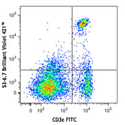
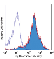
Brilliant Violet 421™ anti-mouse CD279 (PD-1)

Con-A and IL-2 stimulated C57BL/6 splenocytes (3 days) were ... 

Mice were injected subcutaneously with sheep red blood cells... -
Brilliant Violet 605™ anti-mouse CD279 (PD-1)

Con-A and IL-2 stimulated C57BL/6 splenocytes (3 days) were ... -
APC/Cyanine7 anti-mouse CD279 (PD-1)

Con-A and IL-2 stimulated splenocytes (3 days) were stained ... -
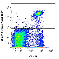
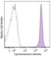
Brilliant Violet 785™ anti-mouse CD279 (PD-1)

Con-A and IL-2 stimulated C57BL/6 splenocytes (3 days) were ... 
-
PE/Dazzle™ 594 anti-mouse CD279 (PD-1)

Con-A and IL-2 stimulated C57BL/6 splenocytes (three days) w... 
-
Alexa Fluor® 647 anti-mouse CD279 (PD-1)

Con A and IL-2 stimulated C57BL/6 splenocytes (three days) w... 
-
Brilliant Violet 711™ anti-mouse CD279 (PD-1)

Con-A and IL-2 stimulated C57BL/6 splenocytes (three days) w... 
-
GoInVivo™ Purified anti-mouse CD279 (PD-1)
-
APC/Fire™ 750 anti-mouse CD279 (PD-1)

Con-A and IL-2 stimulated C57BL/6 mouse splenocytes (3 days)... 
-
Brilliant Violet 510™ anti-mouse CD279 (PD-1)

Con-A and IL-2 stimulated C57BL/6 mouse splenocytes (3 days)... 
-
Ultra-LEAF™ Purified anti-mouse CD279 (PD-1)
Con A and IL-2 stimulated mouse splenocytes (day 3) were sta... -
APC/Fire™ 810 anti-mouse CD279 (PD-1) Antibody

Con-A and IL-2 stimulated C57BL/6 splenocytes (three days) w... -
PE/Fire™ 810 anti-mouse CD279 (PD-1) Antibody

Con-A and IL-2 stimulated C57BL/6 mouse splenocytes (three d... -
PE/Cyanine5 anti-mouse CD279 (PD-1)

Con-A and IL-2 stimulated C57BL/6 splenocytes (3 days) were ... -
PE/Fire™ 640 anti-mouse CD279 (PD-1)

C57BL/6 mouse splenocytes were stimulated with Con-A and IL-... -
Spark Red™ 718 anti-mouse CD279 (PD-1)

Con-A and IL-2 stimulated C57 splenocytes (3 days) were stai... -
PerCP/Fire™ 806 anti-mouse CD279 (PD-1)

Con-A and IL-2 stimulated C57BL/6 splenocytes (three days) w... -
Brilliant Violet 750™ anti-mouse CD279 (PD-1)

Con-A and IL-2 stimulated C57BL/6 splenocytes (3 days) stain... -
PerCP/Fire™ 780 anti-mouse CD279 (PD-1)

Con-A and IL-2 stimulated C57BL/6 splenocytes (three days) w... -
PE/Fire™ 700 anti-mouse CD279 (PD-1)

Unstimulated BALB/c splenocytes were stained with anti-mouse... 
Con A and IL-2 stimulated BALB/c splenocytes (three days) we...


Follow Us