 Login / Register


- Clone
- L291H4 (See other available formats)
- Regulatory Status
- RUO
- Other Names
- CKR4, K5-5, CMKBR4, ChemR13, CC-CKR-4, MGC88293, HGCN:14099
- Isotype
- Mouse IgG1, κ
- Ave. Rating
- Submit a Review
- Product Citations
- publications

-

Human peripheral blood lymphocytes were stained with CD3 FITC and CD194 (clone L291H4) Brilliant Violet 510™ (top) or mouse IgG1, κ Brilliant Violet 510™ isotype control (bottom). -

| Cat # | Size | Price | Quantity Check Availability | Save | ||
|---|---|---|---|---|---|---|
| 359415 | 25 tests | 146€ | ||||
| 359416 | 100 tests | 322€ | ||||
CD194, also known as CCR4, is a CC chemokine receptor. It binds CCL17 and CCL22 and is expressed on a subset of T and B cells, basophils, monocytes, and NK cells. Human Th2 cells are characterized by the expression of CCR4 and CCR8, and these receptors are regulated differently during Th2 development. Human peripheral blood Tregs can be divided into two distinct populations based on the expression of CCR4. Freshly isolated Tregs express CCR4 and presumably represent memory-type Tregs, and CCR4- Tregs require CD3-mediated activation to acquire a regulatory activity. Depletion of CCR4+ T cells leads to Th1-type polarization of CD4+ T cells and augmentation of CD8+ T cell responses to tumor antigens. CCR4 and its ligands are important for the recruitment of memory T cells into the skin in various cutaneous immune diseases.
Product DetailsProduct Details
- Verified Reactivity
- Human
- Reported Reactivity
- Cynomolgus
- Antibody Type
- Monoclonal
- Host Species
- Mouse
- Immunogen
- Human CCR4 transfected cells
- Formulation
- Phosphate-buffered solution, pH 7.2, containing 0.09% sodium azide and BSA (origin USA).
- Preparation
- The antibody was purified by affinity chromatography and conjugated with Brilliant Violet 510™ under optimal conditions.
- Concentration
- Lot-specific (to obtain lot-specific concentration and expiration, please enter the lot number in our Certificate of Analysis online tool.)
- Storage & Handling
- The antibody solution should be stored undiluted between 2°C and 8°C, and protected from prolonged exposure to light. Do not freeze.
- Application
-
FC - Quality tested
- Recommended Usage
-
Each lot of this antibody is quality control tested by immunofluorescent staining with flow cytometric analysis. For flow cytometric staining, the suggested use of this reagent is 5 µl per million cells in 100 µl staining volume or 5 µl per 100 µl of whole blood.
Brilliant Violet 510™ excites at 405 nm and emits at 510 nm. The bandpass filter 510/50 nm is recommended for detection, although filter optimization may be required depending on other fluorophores used. Be sure to verify that your cytometer configuration and software setup are appropriate for detecting this channel. Refer to your instrument manual or manufacturer for support. Brilliant Violet 510™ is a trademark of Sirigen Group Ltd.
Learn more about Brilliant Violet™.
This product is subject to proprietary rights of Sirigen Inc. and is made and sold under license from Sirigen Inc. The purchase of this product conveys to the buyer a non-transferable right to use the purchased product for research purposes only. This product may not be resold or incorporated in any manner into another product for resale. Any use for therapeutics or diagnostics is strictly prohibited. This product is covered by U.S. Patent(s), pending patent applications and foreign equivalents. - Excitation Laser
-
Violet Laser (405 nm)
- Product Citations
-
- RRID
-
AB_2562436 (BioLegend Cat. No. 359415)
AB_2562437 (BioLegend Cat. No. 359416)
Antigen Details
- Structure
- GPCR, seven transmembrane receptor
- Distribution
-
Expressed on a subset of T and B cells, basophils, monocytes, NK cells
- Function
- Migration of inflammatory and regulatory cells to the target tissues
- Ligand/Receptor
- CCL17 and CCL22
- Cell Type
- B cells, Basophils, Embryonic Stem Cells, Monocytes, NK cells, T cells, Tregs
- Biology Area
- Immunology, Innate Immunity, Stem Cells
- Molecular Family
- CD Molecules, Cytokine/Chemokine Receptors, GPCR
- Antigen References
-
1. Katschke KJ, et al. 2001 Arthritis Rheum. 44:1022.
2. Colantonio L, et al. 2002 Eur. J. Immunol. 32:1264.
3. Jakubzick C et al. 2004 Am. J. Pathol. 165:1211.
4. Morimoto Y, et al. 2005 J. Leukoc. Biol. 78:753.
5. Baatar J, et al. 2007 J. Immunol. 178:4891.
6. Kusumoto M, et al. 2007 J. Interferon. Cytokine Res. 27:901. - Gene ID
- 1233 View all products for this Gene ID
- UniProt
- View information about CD194 on UniProt.org
Related Pages & Pathways
Pathways


Related FAQs
- Does staining at room temperature or even at 37°C help for checking chemokine receptors expression?
-
Due to continuous recycling of many chemokine receptors, it may be worthwhile to consider staining at room temperature or at 37°C if the staining at lower temperature (which can potentially reduce receptor turnover) is not optimal.
Other Formats
View All CD194 Reagents Request Custom ConjugationCustomers Also Purchased
Compare Data Across All Formats
This data display is provided for general comparisons between formats.
Your actual data may vary due to variations in samples, target cells, instruments and their settings, staining conditions, and other factors.
If you need assistance with selecting the best format contact our expert technical support team.
-
PerCP/Cyanine5.5 anti-human CD194 (CCR4)

Human peripheral blood lymphocytes were stained with CD3 FIT... -
Purified anti-human CD194 (CCR4)

Human peripheral blood lymphocytes were stained with purifie... 
-
Alexa Fluor® 647 anti-human CD194 (CCR4)

Human peripheral blood lymphocytes were stained with CD3 PE ... 
-
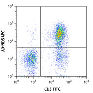
APC anti-human CD194 (CCR4)

Human peripheral blood lymphocytes were stained with CD3 FIT... 
-
PE/Cyanine7 anti-human CD194 (CCR4)

Human peripheral blood lymphocytes were stained with CD3 FIT... -
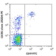
PE anti-human CD194 (CCR4)

Human peripheral blood lymphocytes were stained with CD3 FIT... 
-
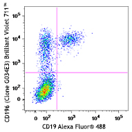
Brilliant Violet 421™ anti-human CD194 (CCR4)

Human peripheral blood lymphocytes were stained with CD3 FIT... 
-
Brilliant Violet 510™ anti-human CD194 (CCR4)

Human peripheral blood lymphocytes were stained with CD3 FIT... 
-
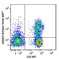
Brilliant Violet 605™ anti-human CD194 (CCR4)

Human peripheral blood lymphocytes were stained with CD3 FIT... 
-
PE/Dazzle™ 594 anti-human CD194 (CCR4)

Human peripheral blood lymphocytes were stained with CD3 APC... 
-
Biotin anti-human CD194 (CCR4)

Human peripheral blood lymphocytes were stained with CD3 APC... -
TotalSeq™-A0071 anti-human CD194 (CCR4)
-
TotalSeq™-C0071 anti-human CD194 (CCR4)
-
TotalSeq™-B0071 anti-human CD194 (CCR4)
-
APC/Fire™ 750 anti-human CD194 (CCR4) Antibody

Human peripheral blood lymphocytes were stained with CD3 FIT... -
PE/Fire™ 810 anti-human CD194 (CCR4) Antibody

Human peripheral blood lymphocytes were stained with anti-hu... -
PE/Fire™ 700 anti-human CD194 (CCR4)

Human peripheral blood lymphocytes were stained with anti-hu... -
TotalSeq™-D0071 anti-human CD194 (CCR4)
-
KIRAVIA Blue 520™ anti-human CD194 (CCR4)

Human peripheral blood lymphocytes were stained with anti-hu... -
APC/Fire™ 810 anti-human CD194 (CCR4)

Human peripheral blood lymphocytes were stained with anti-hu... -
PerCP/Fire™ 780 anti-human CD194 (CCR4)

Human peripheral blood lymphocytes were stained with anti-hu... -
Alexa Fluor® 700 anti-human CD194 (CCR4)

Human peripheral blood lymphocytes were stained with anti-hu... -
PerCP/Fire™ 806 anti-human CD194 (CCR4)

Human peripheral blood lymphocytes were stained with anti-hu... -
Spark Red™ 718 anti-human CD194 (CCR4) (Flexi-Fluor™)


Follow Us