 Login / Register


- Clone
- CC2C6 (See other available formats)
- Regulatory Status
- RUO
- Other Names
- Rh-associated protein, gp42, integrin-associated protein, IAP, neurophilin
- Isotype
- Mouse IgG1, κ
- Ave. Rating
- Submit a Review
- Product Citations
- publications

-

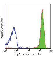
Human peripheral blood lymphocytes stained with biotinylated CC2C6, followed by Sav-PE
| Cat # | Size | Price | Quantity Check Availability | Save | ||
|---|---|---|---|---|---|---|
| 323104 | 100 µg | 169€ | ||||
CD47 also known as Rh-associated protein, gp42, integrin-associated protein (IAP), and neurophilin, is a 42-52 kD member of the immunoglobulin superfamily containing a five-pass transmembrane attachment. Two splice variants have been described in the cytoplasmic tail, the shorter form is expressed in bone-marrow-derived cells, endothelial cells, and fibroblasts while the longer form is expressed by neural tissues. CD47 expression is widely distributed in hematopoietic cells including thymocytes, T cells, B cells, monocytes, platelets, and erythrocytes as well as epithelial cells, endothelial cells, fibroblasts, and neural tissues. CD47 functions as an adhesion molecule and thrombospondin receptor and is non-covalently associated with β3 integrins CD51/CD61, CD41/CD61. Thrombospondin is a ligand for CD47; in the absence of CD47 mice show defects in host defense and β3 integrin-dependent ligand binding, migration, and cellular activation. CD47 is also part of the Rh complex on erythrocytes. The CC2C6 antibody recognizes human CD47 and has been shown to be useful for flow cytometry.
Product DetailsProduct Details
- Verified Reactivity
- Human
- Reported Reactivity
- African Green, Baboon, Cynomolgus, Rhesus
- Antibody Type
- Monoclonal
- Host Species
- Mouse
- Immunogen
- CCRF-CEM T-cell line
- Formulation
- Phosphate-buffered solution, pH 7.2, containing 0.09% sodium azide.
- Preparation
- The antibody was purified by affinity chromatography, and conjugated with biotin under optimal conditions.
- Concentration
- 0.5 mg/ml
- Storage & Handling
- The antibody solution should be stored undiluted between 2°C and 8°C. Do not freeze.
- Application
-
FC - Quality tested
- Recommended Usage
-
Each lot of this antibody is quality control tested by immunofluorescent staining with flow cytometric analysis. For flow cytometric staining, the suggested use of this reagent is ≤2.0 µg per million cells in 100 µl volume. It is recommended that the reagent be titrated for optimal performance for each application.
- Application Notes
-
The CC2C6 monoclonal antibody can block the binding of HCD47 antibody to CD47.
Additional reported applications (for the relevant formats) include: blocking2 - Application References
-
- Seiffert M, et al. 1999. Blood 94:3633.
- Leclair P, et al. 2018. Cell Death Dis. 5:544 (Block)
- Product Citations
-
- RRID
-
AB_756134 (BioLegend Cat. No. 323104)
Antigen Details
- Structure
- Member of the immunoglobulin superfamily, 42-52 kD protein with a five-pass transmembrane attachment. Two splice variants have been described in the cytoplasmic tail.
- Distribution
-
Wide distribution in hematopoietic cells including thymocytes, T cells, B cells, platelets, and erythrocytes as well as epithelial cells, endothelial cells, fibroblasts, and neural tissues.
- Function
- Adhesion molecule and thrombospondin receptor. In the absence of CD47, mice show defects in host defense and beta 3 integrin-dependent ligand binding, migration, and cellular activation. CD47 is a part of the Rh complex on erythrocytes.
- Ligand/Receptor
- Non-covalently associated with ß3 integrins CD51/CD61, CD41/CD61. Thrombospondin is a ligand for CD47.
- Cell Type
- B cells, Endothelial cells, Epithelial cells, Erythrocytes, Fibroblasts, Platelets, T cells, Thymocytes
- Biology Area
- Immunology
- Molecular Family
- CD Molecules
- Antigen References
-
1. Anstee DJ, et al. 1995. In Leucocyte Typing V (Schlossman ed.) Oxford University Press Oxford pp233-234.
2. Brown E, et al. 1990. J. Cell Biol. 111:2785.
3. Gao AG, et al. 1996. J. Biol. Chem. 271:21.
4. Lindberg FP, et al. 1994. J. Biol. Chem. 269:1567. - Gene ID
- 961 View all products for this Gene ID
- UniProt
- View information about CD47 on UniProt.org
Related Pages & Pathways
Pathways


Related FAQs
- How many biotin molecules are per antibody structure?
- We don't routinely measure the number of biotins with our antibody products but the number of biotin molecules range from 3-6 molecules per antibody.
Other Formats
View All CD47 Reagents Request Custom Conjugation| Description | Clone | Applications |
|---|---|---|
| Purified anti-human CD47 | CC2C6 | FC,ICC |
| Biotin anti-human CD47 | CC2C6 | FC |
| FITC anti-human CD47 | CC2C6 | FC |
| PE anti-human CD47 | CC2C6 | FC |
| PerCP/Cyanine5.5 anti-human CD47 | CC2C6 | FC |
| PE/Cyanine7 anti-human CD47 | CC2C6 | FC |
| APC/Fire™ 750 anti-human CD47 | CC2C6 | FC |
| Alexa Fluor® 647 anti-human CD47 | CC2C6 | FC |
| Alexa Fluor® 700 anti-human CD47 | CC2C6 | FC |
| APC anti-human CD47 | CC2C6 | FC |
| Pacific Blue™ anti-human CD47 | CC2C6 | FC |
| Brilliant Violet 421™ anti-human CD47 | CC2C6 | FC |
| Brilliant Violet 605™ anti-human CD47 | CC2C6 | FC |
| TotalSeq™-A0026 anti-human CD47 | CC2C6 | PG |
| TotalSeq™-C0026 anti-human CD47 | CC2C6 | PG |
| PE/Dazzle™ 594 anti-human CD47 Antibody | CC2C6 | FC |
| TotalSeq™-B0026 anti-human CD47 Antibody | CC2C6 | PG |
| TotalSeq™-D0026 anti-human CD47 | CC2C6 | PG |
Customers Also Purchased
Compare Data Across All Formats
This data display is provided for general comparisons between formats.
Your actual data may vary due to variations in samples, target cells, instruments and their settings, staining conditions, and other factors.
If you need assistance with selecting the best format contact our expert technical support team.
-
Purified anti-human CD47

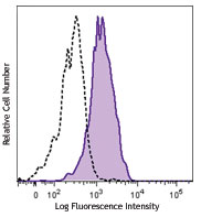
Human peripheral blood lymphocytes stained with purified CC2... 
MCF-7 breast cancer cell line was stained with anti-human CD... -
Biotin anti-human CD47

Human peripheral blood lymphocytes stained with biotinylated... -
FITC anti-human CD47

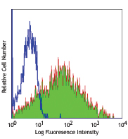
Human peripheral blood lymphocytes stained with CC2C6 FITC -
PE anti-human CD47

Human peripheral blood lymphocytes stained with CC2C6 PE -
PerCP/Cyanine5.5 anti-human CD47

Human peripheral blood lymphocytes were stained with CD47 (c... -
PE/Cyanine7 anti-human CD47

Human peripheral blood monocytes were stained with CD47 (clo... -
APC/Fire™ 750 anti-human CD47

Human peripheral blood monocytes were stained with CD47 (clo... -
Alexa Fluor® 647 anti-human CD47

Human peripheral blood monocytes were stained with CD47 (clo... -
Alexa Fluor® 700 anti-human CD47

Human peripheral blood lymphocytes were stained with CD47 (c... -
APC anti-human CD47

Human peripheral blood lymphocytes were stained with CD47 (c... -
Pacific Blue™ anti-human CD47

Human peripheral blood lymphocytes were stained with CD47 (c... -
Brilliant Violet 421™ anti-human CD47

Human peripheral blood monocytes were stained with CD47 (cl... -
Brilliant Violet 605™ anti-human CD47

Human peripheral blood monocytes were stained with CD47 (clo... -
TotalSeq™-A0026 anti-human CD47
-
TotalSeq™-C0026 anti-human CD47
-
PE/Dazzle™ 594 anti-human CD47 Antibody

Human peripheral blood lymphocytes were stained with anti-hu... -
TotalSeq™-B0026 anti-human CD47 Antibody
-
TotalSeq™-D0026 anti-human CD47


Follow Us