 Login / Register


- Clone
- HM48-1 (See other available formats)
- Regulatory Status
- RUO
- Other Names
- BCM1, SLAMF2, Blast-1
- Isotype
- Armenian Hamster IgG
- Ave. Rating
- Submit a Review
- Product Citations
- publications

-

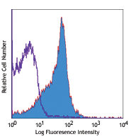
C57BL/6 mouse splenocytes were stained with CD48 (clone HM48-1) APC/Cyanine7 (filled histogram) or Armenian hamster IgG APC/Cyanine7 isotype control (open histogram).
| Cat # | Size | Price | Quantity Check Availability | Save | ||
|---|---|---|---|---|---|---|
| 103431 | 25 µg | 104€ | ||||
| 103432 | 100 µg | 278€ | ||||
CD48 is a 45 kD GPI-anchored glycoprotein also known as BCM1, Blast-1 (human), and OX-45 (rat). It is a member of the Ig superfamily, expressed on T and B cells and monocytes/macrophages. It plays a role in adhesion and T cell recognition. The primary ligands for CD48 are CD2 and CD244.
Product DetailsProduct Details
- Verified Reactivity
- Mouse
- Antibody Type
- Monoclonal
- Host Species
- Armenian Hamster
- Immunogen
- Mouse T lymphoma MBL-2
- Formulation
- Phosphate-buffered solution, pH 7.2, containing 0.09% sodium azide.
- Preparation
- The antibody was purified by affinity chromatography and conjugated with APC/Cyanine7 under optimal conditions.
- Storage & Handling
- The antibody solution should be stored undiluted between 2°C and 8°C, and protected from prolonged exposure to light. Do not freeze.
- Application
-
FC - Quality tested
- Recommended Usage
-
Each lot of this antibody is quality control tested by immunofluorescent staining with flow cytometric analysis. For flow cytometric staining, the suggested use of this reagent is ≤0.25 µg per million cells in 100 µl volume. It is recommended that the reagent be titrated for optimal performance for each application.
- Excitation Laser
-
Red Laser (633 nm)
- Application Notes
-
The HM48-1 antibody is useful for blocking in vitro and in vivo CD48 mediated interactions. Additional reported applications (for the relevant formats) include: immunoprecipitation1, costimulation of T cell proliferation1,2, blocking of CD48-CD2 interaction1, and inhibition of CTL activity and graft rejection1,2. For in vivo studies or highly sensitive assays, we recommend Ultra-LEAF™ purified antibody (Cat. Nos. 103452-103456) with a lower endotoxin limit (Endotoxin < 0.01 EU/µg).
- Additional Product Notes
- BioLegend is in the process of converting the name APC/Cy7 to APC/Cyanine7. The dye molecule remains the same, so you should expect the same quality and performance from our APC/Cyanine7 products. Please contact Technical Service if you have any questions.
- Application References
-
- Kato K, et al. 1992. J. Exp. Med. 176:1241. (IP, Costim, Block)
- Qin L, et al. 1994. J. Exp. Med. 179:341. (Costim, Block)
- Product Citations
-
- RRID
-
AB_2561462 (BioLegend Cat. No. 103431)
AB_2561463 (BioLegend Cat. No. 103432)
Antigen Details
- Structure
- Ig superfamily, 45 kD
- Distribution
-
T cells and B cells, monocytes/macrophages
- Function
- Adhesion, T cell recognition
- Ligand/Receptor
- CD2, CD244
- Cell Type
- B cells, Macrophages, Monocytes, T cells
- Biology Area
- Immunology
- Molecular Family
- Adhesion Molecules, CD Molecules
- Antigen References
-
1. Barclay AN, et al. 1997. The Leukocyte Antigen FactsBook Academic Press.
2. Flament C, et al. 1996. Hum. Immunol. 46:82.
3. Van der Merwe PA, et al. 1995. Curr. Biol. 5:74.
4. Latchman Y, et al. 1998. J. Immunol. 161:5809. - Gene ID
- 12506 View all products for this Gene ID
- UniProt
- View information about CD48 on UniProt.org
Related Pages & Pathways
Pathways


Related FAQs
Other Formats
View All CD48 Reagents Request Custom ConjugationCustomers Also Purchased

Compare Data Across All Formats
This data display is provided for general comparisons between formats.
Your actual data may vary due to variations in samples, target cells, instruments and their settings, staining conditions, and other factors.
If you need assistance with selecting the best format contact our expert technical support team.
-
FITC anti-mouse CD48

C57BL/6 splenocytes stained with HM48-1 FITC -
PE anti-mouse CD48

C57BL/6 mouse splenocytes stained with HM48-1 PE -
Purified anti-mouse CD48

C57BL/6 mouse splenocytes stained with purified HM48-1, foll... 
C57BL/6 mouse frozen spleen section was fixed with 4% parafo... -
Biotin anti-mouse CD48

C57BL/6 mouse splenocytes stained with biotinylated HM48-1, ... -
APC anti-mouse CD48

C57BL/6 mouse splenocytes stained with HM48-1 APC -
Alexa Fluor® 488 anti-mouse CD48

C57BL/6 mouse splenocytes stained with HM48-1 Alexa Fluor&re... -
Alexa Fluor® 647 anti-mouse CD48

C57BL/6 mouse splenocytes stained with HM48-1 Alexa Fluor&re... -
Pacific Blue™ anti-mouse CD48

C57BL/6 mouse splenocytes stained with Pacific blue™ H... -
PE/Cyanine5 anti-mouse CD48

C57BL/6 splenocytes stained with HM48-1 PE/Cyanine5 -
PerCP/Cyanine5.5 anti-mouse CD48

C57BL/6 mouse splenocytes were stained with CD48 (clone HM48... -
PE/Cyanine7 anti-mouse CD48

C57BL/6 splenocytes stained with HM48-1 PE/Cyanine7 -
Alexa Fluor® 700 anti-mouse CD48

C57BL/6 splenocytes stained with HM48-1 Alexa Fluor® 700 -
Brilliant Violet 421™ anti-mouse CD48

C57BL/6 mouse splenocytes were stained with CD48 (clone HM48... -
APC/Cyanine7 anti-mouse CD48

C57BL/6 mouse splenocytes were stained with CD48 (clone HM48... -
Purified anti-mouse CD48 (Maxpar® Ready)

C57BL/6 mouse splenocytes stained with 176Yb-anti-CD45R/B220... -
Brilliant Violet 605™ anti-mouse CD48

C57BL/6 mouse splenocytes were stained with CD48 (clone HM48... -
Brilliant Violet 510™ anti-mouse CD48

C57BL/6 mouse splenocytes were stained with CD48 (clone HM48... -
Brilliant Violet 711™ anti-mouse CD48

C57BL/6 mouse splenocytes were stained with CD48 (clone HM48... -
PE/Dazzle™ 594 anti-mouse CD48

C57BL/6 mouse splenocytes were stained with CD48 (clone HM48... -
APC/Fire™ 750 anti-mouse CD48

C57BL/6 mouse splenocytes were stained with CD48 (clone HM48... -
TotalSeq™-A0429 anti-mouse CD48
-
Brilliant Violet 785™ anti-mouse CD48

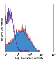
C57BL/6 splenocytes were stained with CD48 (clone HM48-1) Br... -
Ultra-LEAF™ Purified anti-mouse CD48
C57BL/6 mouse splenocytes stained with Ultra-LEAF™ purified ... -
TotalSeq™-B0429 anti-mouse CD48
-
TotalSeq™-C0429 anti-mouse CD48


Follow Us