 Login / Register


- Clone
- MIH18 (See other available formats)
- Regulatory Status
- RUO
- Workshop
- HCDM listed
- Other Names
- B7-DC, PD-L2, PDL2, B7DC
- Isotype
- Mouse IgG1, κ
- Ave. Rating
- Submit a Review
- Product Citations
- publications

-

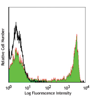
Human monocyte-derived dendritic cells were stained with anti-human CD273 (B7-DC, PD-L2) (clone MIH18) APC/Cyanine7 (filled histogram) or mouse IgG1, κ APC/Cyanine7 isotype control (open histogram).
| Cat # | Size | Price | Quantity Check Availability | Save | ||
|---|---|---|---|---|---|---|
| 345515 | 25 tests | 123€ | ||||
| 345516 | 100 tests | 300€ | ||||
CD273, known as B7-DC, is also called programmed death ligand 2 (PDL2). This ligand is a 25 kD type I transmembrane protein and a member of B7 family within the immunoglobulin receptor superfamily and is expressed on a subset of dendritic cells, liver and a small subset of macrophages as well as a few transformed cell lines. CD273 has been reported to be stimulatory on dendritic cells when cross-linked and to inhibit T cell activation upon engaging the PD-1 receptor. CD273 has also been reported to bind to an alternative receptor and to mediate T cell activation through such non-PD1 mediated interactions. Clone MIH18 is reported to block PDL2.
Product DetailsProduct Details
- Verified Reactivity
- Human
- Antibody Type
- Monoclonal
- Host Species
- Mouse
- Immunogen
- Human B7-DC transfected cells
- Formulation
- Phosphate-buffered solution, pH 7.2, containing 0.09% sodium azide and BSA (origin USA)
- Preparation
- The antibody was purified by affinity chromatography and conjugated with APC/Cyanine7 under optimal conditions.
- Concentration
- Lot-specific (to obtain lot-specific concentration and expiration, please enter the lot number in our Certificate of Analysis online tool.)
- Storage & Handling
- The antibody solution should be stored undiluted between 2°C and 8°C, and protected from prolonged exposure to light. Do not freeze.
- Application
-
FC - Quality tested
- Recommended Usage
-
Each lot of this antibody is quality control tested by immunofluorescent staining with flow cytometric analysis. For flow cytometric staining, the suggested use of this reagent is 5 µl per million cells or 5 µl per 100 µl of whole blood. It is recommended that the reagent be titrated for optimal performance for each application.
- Excitation Laser
-
Red Laser (633 nm)
- Application Notes
-
Additional reported applications (for the relevant formats) include: blocking4,5, and immunohistochemistry in frozen sections2 and paraffin-embedded formalin-fixed sections6. The LEAF™ purified antibody (Endotoxin <0.1 EU/µg, Azide-free, 0.2 µm filtered) is recommended for functional assays (Cat. No. 345504).
- Additional Product Notes
-
BioLegend is in the process of converting the name APC/Cy7 to APC/Cyanine7. The dye molecule remains the same, so you should expect the same quality and performance from our APC/Cyanine7 products. Please contact Technical Service if you have any questions.
- Application References
-
- Youngnak-Piboonratankit P, et al. 2004. Immunol. Lett. 94:215. (IHC, IF)
- Ohigashi Y, et al. 2005. Clin. Cancer. Res. 8:2947. (IHC, IF)
- Hobo W, et al. 2010. Blood 25:4501. (FC)
- Nagamatsu T, et al. 2009. Hum. Reprod. 24:3160. (Block)
- Alvarez IB, et al. 2010. J. Infect. Dis. 202:524. (Block)
- Taube JM, et al. 2014. Clin. Cancer Res. 19:5064. (IHC) PubMed
- RRID
-
AB_2783233 (BioLegend Cat. No. 345515)
AB_2783234 (BioLegend Cat. No. 345516)
Antigen Details
- Structure
- B7 Immunoglobulin superfamily, 25 kD
- Distribution
-
Dendritic cells, liver, few transformed cell lines, subset of macrophages
- Function
- Binds to PD-1 and alternative receptor; ligation on DC stimulates, inhibits T cell responses via PD-1 binding, stimulates T cells via alternative receptor binding and promotes tumor immunity
- Ligand/Receptor
- PD-1
- Cell Type
- Dendritic cells, Macrophages
- Biology Area
- Costimulatory Molecules, Immunology
- Molecular Family
- CD Molecules, Immune Checkpoint Receptors
- Antigen References
-
1. Carreno BM, et al. 2002. Annu. Rev. Immunol. 20:29.
2. Ohigashi Y, et al. 2005. Clin. Cancer. Res. 8:2947. - Gene ID
- 80380 View all products for this Gene ID
- UniProt
- View information about CD273 on UniProt.org
Related Pages & Pathways
Pathways


Related FAQs
Other Formats
View All CD273 Reagents Request Custom Conjugation| Description | Clone | Applications |
|---|---|---|
| Purified anti-human CD273 (B7-DC, PD-L2) | MIH18 | FC,IHC-F,IHC-P,Block |
| PE anti-human CD273 (B7-DC, PD-L2) | MIH18 | FC |
| APC anti-human CD273 (B7-DC, PD-L2) | MIH18 | FC |
| Biotin anti-human CD273 (B7-DC, PD-L2) | MIH18 | FC |
| PE/Cyanine7 anti-human CD273 (B7-DC, PD-L2) | MIH18 | FC |
| Alexa Fluor® 647 anti-human CD273 (B7-DC, PD-L2) | MIH18 | FC |
| APC/Cyanine7 anti-human CD273 (B7-DC, PD-L2) | MIH18 | FC |
| PE/Dazzle™ 594 anti-human CD273 (B7-DC, PD-L2) | MIH18 | FC |
| Brilliant Violet 421™ anti-human CD273 (B7-DC, PD-L2) | MIH18 | FC |
| Ultra-LEAF™ Purified anti-human CD273 (B7-DC, PD-L2) | MIH18 | FC,IHC-F,IHC-P,Block |
| Brilliant Violet 650™ anti-human CD273 (B7-DC, PD-L2) | MIH18 | FC |
| Brilliant Violet 750™ anti-human CD273 (B7-DC, PD-L2) | MIH18 | FC |
Customers Also Purchased
Compare Data Across All Formats
This data display is provided for general comparisons between formats.
Your actual data may vary due to variations in samples, target cells, instruments and their settings, staining conditions, and other factors.
If you need assistance with selecting the best format contact our expert technical support team.
-
Purified anti-human CD273 (B7-DC, PD-L2)

Human monocyte-derived dendritic cells were stained with CD2... -
PE anti-human CD273 (B7-DC, PD-L2)

Human monocyte-derived dendritic cells were stained with ant... -
APC anti-human CD273 (B7-DC, PD-L2)

Human monocyte-derived dendritic cells were stained with ant... -
Biotin anti-human CD273 (B7-DC, PD-L2)

Human monocyte-derived dendritic cells were stained with bio... -
PE/Cyanine7 anti-human CD273 (B7-DC, PD-L2)

Human monocyte-derived dendritic cells were stained with ant... -
Alexa Fluor® 647 anti-human CD273 (B7-DC, PD-L2)

Human monocyte-derived dendritic cells were stained with CD2... -
APC/Cyanine7 anti-human CD273 (B7-DC, PD-L2)

Human monocyte-derived dendritic cells were stained with ant... -
PE/Dazzle™ 594 anti-human CD273 (B7-DC, PD-L2)

Human monocyte-derived dendritic cells were stained with ant... -
Brilliant Violet 421™ anti-human CD273 (B7-DC, PD-L2)

Human monocyte-derived dendritic cells were stained with ant... -
Ultra-LEAF™ Purified anti-human CD273 (B7-DC, PD-L2)
Human monocyte-derived dendritic cells were stained with CD2... -
Brilliant Violet 650™ anti-human CD273 (B7-DC, PD-L2)

Human monocyte-derived dendritic cells were stained with ant... -
Brilliant Violet 750™ anti-human CD273 (B7-DC, PD-L2)

Human monocyte-derived dendritic cells were stained with ant...


Follow Us