 Login / Register


- Clone
- NKI-M9 (See other available formats)
- Regulatory Status
- RUO
- Workshop
- IV 103
- Other Names
- Vitronectin receptor α chain, Integrin αV, αV integrin, ITGAV
- Isotype
- Mouse IgG2a, κ
- Ave. Rating
- Submit a Review
- Product Citations
- publications

-

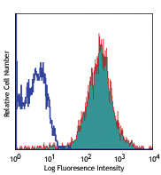
Human melanoma cell line M21 was stained with CD51 (clone NKI-M9) APC (filled histogram) or mouse IgG2a, κ APC isotype control (open histogram).
| Cat # | Size | Price | Quantity Check Availability | Save | ||
|---|---|---|---|---|---|---|
| 327913 | 25 tests | 76€ | ||||
| 327914 | 100 tests | 180€ | ||||
CD51 is a type I integral membrane glycoprotein, known as vitronectin receptor α chain, or integrin αV. It forms heterodimer with integrin β1 (CD29), β3 (CD61), β5, β6, or β8. CD51 contains two disulfide-linked subunits of 125 kD and 24 kD, and is expressed on endothelial cells, fibroblasts, macrophages, platelets, osteoclasts, neuroblastoma, melanoma, and hepatoma cells. Many extracellular matrix proteins with RGD-motifs are CD51 ligands. In association with its β chains, CD51 binds vitronectin, von Willebrand factor, fibronectin, thrombospondin, osteopontin, fibrinogen, and laminin. CD51, as an adhesion molecule, plays important roles in leukocytes homing and rolling, mediates bone absorption and angiogenesis.
Product DetailsProduct Details
- Verified Reactivity
- Human
- Antibody Type
- Monoclonal
- Host Species
- Mouse
- Immunogen
- Melonama cells
- Formulation
- Phosphate-buffered solution, pH 7.2, containing 0.09% sodium azide and BSA (origin USA)
- Preparation
- The antibody was purified by affinity chromatography and conjugated with APC under optimal conditions.
- Concentration
- Lot-specific (to obtain lot-specific concentration and expiration, please enter the lot number in our Certificate of Analysis online tool.)
- Storage & Handling
- The antibody solution should be stored undiluted between 2°C and 8°C, and protected from prolonged exposure to light. Do not freeze.
- Application
-
FC - Quality tested
- Recommended Usage
-
Each lot of this antibody is quality control tested by immunofluorescent staining with flow cytometric analysis. For flow cytometric staining, the suggested use of this reagent is 5 µL per million cells in 100 µL staining volume or 5 µL per 100 µL of whole blood. It is recommended that the reagent be titrated for optimal performance for each application.
- Excitation Laser
-
Red Laser (633 nm)
- Application References
-
- Knapp W, et al. Eds. 1989. Leucocyte Typing IV. Oxford University Press. New York.
- Defilippi P, et al. 1991. J. Cell Biol. 114:855. PubMed
- Burdick MM, et al. 2003. Am. J. Physiol.Cell Ph. 284:C977. PubMed
- Grzeszkiewicz TM, et al. 2001. J. Biol. Chem. 276:21943.
- Sonnenberg A, et al. 1990. J. Cell Biol. 110:2145. PubMed
- Silverman AP, et al. 2008. J Mol Biol. 385:1064. PubMed
- Khurana S, et al. 2013. Blood. 121:2587. PubMed
- RRID
-
AB_2876633 (BioLegend Cat. No. 327913)
AB_2876633 (BioLegend Cat. No. 327914)
Antigen Details
- Structure
- Integrin, forms heterodimer with integrin β1 (CD29), β3 (CD61), β5, β6, or β8, 125 kD and 24 kD in reduced condition, 150 kD (unreduced)
- Distribution
-
Endothelial cells, fibroblasts, macrophages, platelets, osteoclasts, neuroblastoma, melanoma, and hepatoma cells.
- Function
- Adhesion, angiogenesis
- Ligand/Receptor
- Vitronectin, von Willebrand factor, fibrinogen, thrombospondin, osteopontin, denatured collagen, fibronectin, laminin.
- Cell Type
- Endothelial cells, Fibroblasts, Macrophages, Mesenchymal Stem Cells, Osteoclasts, Platelets
- Biology Area
- Angiogenesis, Cell Adhesion, Cell Biology, Costimulatory Molecules, Immunology, Stem Cells
- Molecular Family
- CD Molecules
- Antigen References
-
1. Nesbitt S, et al. 1993. J. Biol. Chem. 268:16737.
2. Zola H, et al. 2007. Leukocyte and Stromal Cell Molecules:The CD Markers Wiley-Liss A John Wiley & Sons Inc, Publication - Gene ID
- 3685 View all products for this Gene ID
- UniProt
- View information about CD51 on UniProt.org


Related FAQs
Other Formats
View All CD51 Reagents Request Custom Conjugation| Description | Clone | Applications |
|---|---|---|
| Purified anti-human CD51 | NKI-M9 | FC,ICC,ELISA,Block,IP |
| Biotin anti-human CD51 | NKI-M9 | FC |
| FITC anti-human CD51 | NKI-M9 | FC |
| PE anti-human CD51 | NKI-M9 | FC |
| Ultra-LEAF™ Purified anti-human CD51 | NKI-M9 | FC,ICC,ELISA,Block,IP |
| APC anti-human CD51 | NKI-M9 | FC |
| PE/Cyanine7 anti-human CD51 | NKI-M9 | FC |
Customers Also Purchased
Compare Data Across All Formats
This data display is provided for general comparisons between formats.
Your actual data may vary due to variations in samples, target cells, instruments and their settings, staining conditions, and other factors.
If you need assistance with selecting the best format contact our expert technical support team.
-
Purified anti-human CD51

Human melanoma cell line M21 stained with purified NKI-M9, f... 
MDA-MB231 breast cancer cell line was stained with anti-huma... -
Biotin anti-human CD51

Human melanoma cell line M21 stained with biotinylated NKI-M... -
FITC anti-human CD51

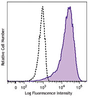
Human melanoma cell line M21 stained with NKI-M9 FITC -
PE anti-human CD51

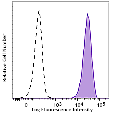
Human melanoma cell line M21 stained with NKI-M9 PE -
Ultra-LEAF™ Purified anti-human CD51

Human melanoma cell line M21 stained with Ultra-LEAF™ purifi...
MDA-MB231 breast cancer cell line was stained with anti-huma... -
APC anti-human CD51

Human melanoma cell line M21 was stained with CD51 (clone NK... -
PE/Cyanine7 anti-human CD51

Human melanoma cell line M21 was stained with CD51 (clone NK...


Follow Us