 Login / Register


- Clone
- HIT2 (See other available formats)
- Regulatory Status
- RUO
- Workshop
- III 155
- Other Names
- T10, ADP-ribosyl cyclase
- Isotype
- Mouse IgG1, κ
- Ave. Rating
- Submit a Review
- Product Citations
- publications

-

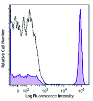
Human peripheral blood lymphocytes stained with HIT2 Alexa Fluor® 700 -

Confocal image of human lymph node sample acquired using the IBEX method of highly multiplexed antibody-based imaging: CD38 (blue) in Cycle 5 and Clec9a (magenta) in Cycle 9. Tissues were prepared using ~1% (vol/vol) formaldehyde and a detergent. Following fixation, samples are immersed in 30% (wt/vol) sucrose for cryoprotection. Images are courtesy of Drs. Andrea J. Radtke and Ronald N. Germain of the Center for Advanced Tissue Imaging (CAT-I) in the National Institute of Allergy and Infectious Diseases (NIAID, NIH).
| Cat # | Size | Price | Quantity Check Availability | Save | ||
|---|---|---|---|---|---|---|
| 303523 | 25 µg | 100€ | ||||
| 303524 | 100 µg | 212€ | ||||
CD38 is a 45 kD type II transmembrane glycoprotein also known as T10. It is an ADP-ribosyl hydrolase expressed at variable levels on hematopoietic cells and in some non-hematopoietic tissues (such as brain, muscles, and kidney). In humans, it is expressed at high levels on plasma cells and activated T and B cells. By functioning as both a cyclase and a hydrolase, CD38 mediates lymphocyte activation, adhesion, and the metabolism of cADPR and NAADP. CD31 is the ligand of CD38.
Product DetailsProduct Details
- Verified Reactivity
- Human
- Reported Reactivity
- Chimpanzee, Horse, Cow
- Antibody Type
- Monoclonal
- Host Species
- Mouse
- Formulation
- Phosphate-buffered solution, pH 7.2, containing 0.09% sodium azide.
- Preparation
- The antibody was purified by affinity chromatography and conjugated with Alexa Fluor® 700 under optimal conditions.
- Concentration
- 0.5 mg/ml
- Storage & Handling
- The antibody solution should be stored undiluted between 2°C and 8°C, and protected from prolonged exposure to light. Do not freeze.
- Application
-
FC - Quality tested
SB - Reported in the literature, not verified in house - Recommended Usage
-
Each lot of this antibody is quality control tested by immunofluorescent staining with flow cytometric analysis. The suggested use of this reagent is ≤ 1.0 µg per 106 cells in 100 µl volume. It is highly recommended that the reagent be titrated for optimal performance for each application.
* Alexa Fluor® 700 has a maximum emission of 719 nm when it is excited at 633nm / 635nm. Prior to using Alexa Fluor® 700 conjugate for flow cytometric analysis, please verify your flow cytometer's capability of exciting and detecting the fluorochrome.
Alexa Fluor® and Pacific Blue™ are trademarks of Life Technologies Corporation.
View full statement regarding label licenses - Excitation Laser
-
Red Laser (633 nm)
- Application Notes
-
Additional reported applications (for the relevant formats) include: immunohistochemical staining of acetone-fixed frozen tissue sections6 and spatial biology (IBEX)10,11.
- Additional Product Notes
-
Iterative Bleaching Extended multi-pleXity (IBEX) is a fluorescent imaging technique capable of highly-multiplexed spatial analysis. The method relies on cyclical bleaching of panels of fluorescent antibodies in order to image and analyze many markers over multiple cycles of staining, imaging, and, bleaching. It is a community-developed open-access method developed by the Center for Advanced Tissue Imaging (CAT-I) in the National Institute of Allergy and Infectious Diseases (NIAID, NIH).
- Application References
-
- Kishimoto T, et al. Eds. 1997. Leucocyte Typing VI. Garland Publishing Inc. London.
- Dieu M. 1998. J. Exp. Med. 188:373.
- Esser M, et al. 2001. J. Virol. 75:6173.
- Jeannin P, et al. 1999. J. Immunol. 162:2044.
- Kapsogeorgou EK, et al. 2001. J. Immunol. 166:3107.
- van der Voort R, et al. 1997. J. Exp. Med. 185:2121. (IHC)
- Bende RJ, et al. 2003. Am. J. Pathol. 162:105.
- Lehner M, et al. 2008. J. Leukoc. Biol. 83:883. PubMed
- Yoshino N, et al. 2000. Exp. Anim. (Tokyo) 49:97. (FC)
- Radtke AJ, et al. 2020. Proc Natl Acad Sci USA. 117:33455-33465. (SB) PubMed
- Radtke AJ, et al. 2022. Nat Protoc. 17:378-401. (SB) PubMed
- Product Citations
-
- RRID
-
AB_2228785 (BioLegend Cat. No. 303523)
AB_2072781 (BioLegend Cat. No. 303524)
Antigen Details
- Structure
- ADP-ribosyl cyclase, ectoenzyme, type II glycoprotein, 45 kD
- Distribution
-
T cells, B cells, NK, myeloid, plasma, and dendritic cells
- Function
- Ecto-ADP-ribosyl cyclase, calcium signaling, cell activation
- Ligand/Receptor
- CD31, hyaluronic acid
- Cell Type
- B cells, Dendritic cells, NK cells, Plasma cells, T cells
- Biology Area
- Immunology
- Molecular Family
- Adhesion Molecules, CD Molecules
- Antigen References
-
1. Ferrero E, et al. 1999. J. Leukoc. Biol. 65:151.
2. Lund F, et al. 1995. Immunol. Today 16:469. - Gene ID
- 952 View all products for this Gene ID
- UniProt
- View information about CD38 on UniProt.org
Related Pages & Pathways
Pathways


Related FAQs
- If an antibody clone has been previously successfully used in IBEX in one fluorescent format, will other antibody formats work as well?
-
It’s likely that other fluorophore conjugates to the same antibody clone will also be compatible with IBEX using the same sample fixation procedure. Ultimately a directly conjugated antibody’s utility in fluorescent imaging and IBEX may be specific to the sample and microscope being used in the experiment. Some antibody clone conjugates may perform better than others due to performance differences in non-specific binding, fluorophore brightness, and other biochemical properties unique to that conjugate.
- Will antibodies my lab is already using for fluorescent or chromogenic IHC work in IBEX?
-
Fundamentally, IBEX as a technique that works much in the same way as single antibody panels or single marker IF/IHC. If you’re already successfully using an antibody clone on a sample of interest, it is likely that clone will have utility in IBEX. It is expected some optimization and testing of different antibody fluorophore conjugates will be required to find a suitable format; however, legacy microscopy techniques like chromogenic IHC on fixed or frozen tissue is an excellent place to start looking for useful antibodies.
- Are other fluorophores compatible with IBEX?
-
Over 18 fluorescent formats have been screened for use in IBEX, however, it is likely that other fluorophores are able to be rapidly bleached in IBEX. If a fluorophore format is already suitable for your imaging platform it can be tested for compatibility in IBEX.
- The same antibody works in one tissue type but not another. What is happening?
-
Differences in tissue properties may impact both the ability of an antibody to bind its target specifically and impact the ability of a specific fluorophore conjugate to overcome the background fluorescent signal in a given tissue. Secondary stains, as well as testing multiple fluorescent conjugates of the same clone, may help to troubleshoot challenging targets or tissues. Using a reference control tissue may also give confidence in the specificity of your staining.
- How can I be sure the staining I’m seeing in my tissue is real?
-
In general, best practices for validating an antibody in traditional chromogenic or fluorescent IHC are applicable to IBEX. Please reference the Nature Methods review on antibody based multiplexed imaging for resources on validating antibodies for IBEX.
Other Formats
View All CD38 Reagents Request Custom ConjugationCustomers Also Purchased
Compare Data Across All Formats
This data display is provided for general comparisons between formats.
Your actual data may vary due to variations in samples, target cells, instruments and their settings, staining conditions, and other factors.
If you need assistance with selecting the best format contact our expert technical support team.
-
APC anti-human CD38

Human peripheral blood lymphocytes stained with HIT2 APC -
FITC anti-human CD38

Human peripheral blood lymphocytes stained with HIT2 FITC -
PE anti-human CD38

Human peripheral blood lymphocytes stained with HIT2 PE -
PE/Cyanine5 anti-human CD38

Human peripheral blood lymphocytes stained with HIT2 PE/Cyan... -
Purified anti-human CD38

Human peripheral blood lymphocytes stained with purified HIT... -
Alexa Fluor® 488 anti-human CD38

Human peripheral blood lymphocytes stained with HIT2 Alexa F... -
Alexa Fluor® 647 anti-human CD38

Human peripheral blood lymphocytes stained with HIT2 Alexa F... -
PE/Cyanine7 anti-human CD38

Human peripheral blood lymphocytes stained with HIT2 PE/Cyan... -
Biotin anti-human CD38

Human peripheral blood lymphocytes stained with biotinylated... -
PerCP anti-human CD38

Human peripheral blood lymphocytes stained with HIT2 PerCP -
PerCP/Cyanine5.5 anti-human CD38

Human peripheral blood lymphocytes were stained with CD38 (c... -
Alexa Fluor® 700 anti-human CD38

Human peripheral blood lymphocytes stained with HIT2 Alexa F... 
Confocal image of human lymph node sample acquired using the... -
Brilliant Violet 421™ anti-human CD38

Human peripheral blood lymphocytes were stained with CD38 (c... -
Brilliant Violet 711™ anti-human CD38

Human peripheral blood lymphocytes were stained with CD38 (c... -
Brilliant Violet 785™ anti-human CD38

Human peripheral blood lymphocytes were stained with CD38 (c... -
Brilliant Violet 605™ anti-human CD38

Human peripheral blood lymphocytes were stained with CD38 (c... -
APC/Cyanine7 anti-human CD38

Human peripheral blood lymphocytes were stained with CD38 (c... -
Purified anti-human CD38 (Maxpar® Ready)

Human PBMCs stained with 154Sm-anti-CD45 (HI30) and 167Er-an... -
PE/Dazzle™ 594 anti-human CD38

Human peripheral blood granulocytes were stained with CD38 (... -
PE anti-human CD38

Typical results from human peripheral blood lymphocytes stai... -
Brilliant Violet 510™ anti-human CD38

Human peripheral blood lymphocytes were stained with CD38 (c... -
FITC anti-human CD38

Typical results from human peripheral blood lymphocytes stai... -
PE/Dazzle™ 594 anti-human CD38
Typical results from human peripheral blood granulocytes sta... -
TotalSeq™-A0389 anti-human CD38
-
TotalSeq™-C0389 anti-human CD38
-
APC/Fire™ 750 anti-human CD38

Human peripheral blood lymphocytes were stained with CD38 (C... -
TotalSeq™-B0389 anti-human CD38
-
APC/Fire™ 810 anti-human CD38

Human peripheral blood lymphocytes were stained with CD38 (c... -
Spark NIR™ 685 anti-human CD38 Antibody

Human peripheral blood lymphocytes were stained with CD38 (c... -
TotalSeq™-D0389 anti-human CD38
-
PerCP/Cyanine5.5 anti-human CD38

Typical results from human peripheral blood lymphocytes stai... -
APC anti-human CD38

Typical results from human peripheral blood lymphocytes stai... -
APC/Fire™ 750 anti-human CD38

Typical results from human peripheral blood lymphocytes stai... -
PE/Cyanine7 anti-human CD38

Typical results from human peripheral blood lymphocytes stai... -
GMP PE anti-human CD38

Typical results from human peripheral blood lymphocytes stai... -
GMP FITC anti-human CD38

Typical results from human peripheral blood lymphocytes stai... -
Pacific Blue™ anti-human CD38

Human peripheral blood lymphocytes were stained with anti-hu... -
Pacific Blue™ anti-human CD38

Typical results from human peripheral blood lymphocytes stai... -
GMP APC/Fire™ 750 anti-human CD38

Typical results from human peripheral blood lymphocytes stai... -
GMP PE/Dazzle™ 594 anti-human CD38

Typical results from human peripheral blood lymphocytes stai... -
GMP APC anti-human CD38

Typical results from human peripheral blood lymphocytes stai... -
GMP PE/Cyanine7 anti-human CD38

Typical results from human peripheral blood lymphocytes stai... -
Spark Blue™ 515 anti-human CD38

Human peripheral blood cells were surface stained with (left... -
GMP Pacific Blue™ anti-human CD38

Typical results from human peripheral blood lymphocytes stai... -
Spark Violet™ 423 anti-human CD38

Human peripheral blood lymphocytes were stained with anti-hu...


Follow Us