 Login / Register


- Clone
- FA-11 (See other available formats)
- Regulatory Status
- RUO
- Other Names
- Macrosialin
- Isotype
- Rat IgG2a
- Ave. Rating
- Submit a Review
- Product Citations
- publications

-

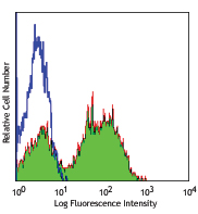
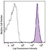
Thioglycolate-elicited Balb/c peritoneal macrophages intracelluar stained with FA-11 Alexa Fluor® 647
| Cat # | Size | Price | Quantity Check Availability | Save | ||
|---|---|---|---|---|---|---|
| 137003 | 25 µg | 118€ | ||||
| 137004 | 100 µg | 278€ | ||||
Mouse CD68, also known as macrosialin, is an 85-115 kD member of the lysosomal-associated membrane protein (LAMP) family. It is a heavily glycosylated and predominantly intracellular protein, mainly in late endosomes. Macrosialin is the murine homolog to the human macrophage glycoprotein CD68. It is expressed on tissue macrophages, Langerhans cells and at low levels on dendritic cells. Lamp proteins may have functions relating to cell-cell interaction or cell-ligand interaction. The biological function of CD68 is not completely understood.
Product DetailsProduct Details
- Verified Reactivity
- Mouse
- Antibody Type
- Monoclonal
- Host Species
- Rat
- Immunogen
- Purified Con A receptor glycoproteins from the P815 cell line
- Formulation
- Phosphate-buffered solution, pH 7.2, containing 0.09% sodium azide.
- Preparation
- The antibody was purified by affinity chromatography and conjugated with Alexa Fluor® 647 under optimal conditions.
- Concentration
- 0.5 mg/ml
- Storage & Handling
- The antibody solution should be stored undiluted between 2°C and 8°C, and protected from prolonged exposure to light. Do not freeze.
- Application
-
ICFC - Quality tested
FC - Verified
SB - Community verified - Recommended Usage
-
Each lot of this antibody is quality control tested by intracellular immunofluorescent staining with flow cytometric analysis. For flow cytometric staining, the suggested use of this reagent is ≤0.06 µg per million cells in 100 µl volume. It is recommended that the reagent be titrated for optimal performance for each application.
* Alexa Fluor® 647 has a maximum emission of 668 nm when it is excited at 633 nm / 635 nm.
Alexa Fluor® and Pacific Blue™ are trademarks of Life Technologies Corporation.
View full statement regarding label licenses - Excitation Laser
-
Red Laser (633 nm)
- Application Notes
-
Additional reported (for relevant formats) applications include: immunoprecipitation1, 2, Western Blot1, 2, immunohistochemical staining of frozen sections2 and paraformaldehyde-fixed paraffin-embedded sections3, and spatial biology (IBEX)9,10.
- Additional Product Notes
-
This product has been verified for IHC-P (Immunohistochemistry - formalin-fixed paraffin-embedded tissues) on the NanoString GeoMx® Digital Spatial Profiler. The GeoMx® enables researchers to perform spatial analysis of protein and RNA targets in FFPE and fresh frozen human and mouse samples. For more information about our spatial biology products and the GeoMx® platform, please visit our spatial biology page.
- Application References
-
- Silva RP, et al. 1999. Biochem. J. 338:687. (IP, WB)
- Rabinowitz SS, et al. 1991. J. Exp. Med. 174:827. (IP, WB, IHC)
- Wu J, et al. 2008. P. Natl. Acad. Sci. USA 105:16934. (IHC)
- Kayama H, et al. 2012. PNAS. 109:5010. PubMed
- Park S, et al. 2013. Biomaterials. 34:598. PubMed
- Guiducci C, et al. 2013. J Exp Med. 210:2903. PubMed
- McKinstry SU, et al. 2014. J Neurosci. 34:9455. PubMed
- Li X, et al. 2015. J Am Heart Assoc. 6:4. PubMed
- Radtke AJ, et al. 2020. Proc Natl Acad Sci U S A. 117:33455-65. (SB) PubMed
- Radtke AJ, et al. 2022. Nat Protoc. 17:378-401. (SB) PubMed
- Product Citations
-
- RRID
-
AB_2044001 (BioLegend Cat. No. 137003)
AB_2044002 (BioLegend Cat. No. 137004)
Antigen Details
- Structure
- A member of the lysosomal-associated membrane protein (lamp) family.
- Distribution
-
Expressed on tissue macrophages, Langerhans cells, and at low levels on dendritic cells.
- Function
- Involved in cell-cell interaction or cell-ligand interaction, still not completely understood.
- Cell Type
- Antigen-presenting cells, Dendritic cells, Langerhans cells, Leukocytes, Macrophages
- Biology Area
- Cell Biology, Immunology, Innate Immunity, Neuroscience, Neuroscience Cell Markers
- Molecular Family
- Adhesion Molecules, CD Molecules, Innate Immune Signaling
- Antigen References
-
1. Ramprasad MP, et al. 1996. Proc. Natl. Acad. Sci. USA 93:14833.
2. Smith MJ, et al. 1987. J. Cell. Sci. 87:113. - Gene ID
- 12514 View all products for this Gene ID
- UniProt
- View information about CD68 on UniProt.org
Related Pages & Pathways
Pathways


Related FAQs
- If an antibody clone has been previously successfully used in IBEX in one fluorescent format, will other antibody formats work as well?
-
It’s likely that other fluorophore conjugates to the same antibody clone will also be compatible with IBEX using the same sample fixation procedure. Ultimately a directly conjugated antibody’s utility in fluorescent imaging and IBEX may be specific to the sample and microscope being used in the experiment. Some antibody clone conjugates may perform better than others due to performance differences in non-specific binding, fluorophore brightness, and other biochemical properties unique to that conjugate.
- Will antibodies my lab is already using for fluorescent or chromogenic IHC work in IBEX?
-
Fundamentally, IBEX as a technique that works much in the same way as single antibody panels or single marker IF/IHC. If you’re already successfully using an antibody clone on a sample of interest, it is likely that clone will have utility in IBEX. It is expected some optimization and testing of different antibody fluorophore conjugates will be required to find a suitable format; however, legacy microscopy techniques like chromogenic IHC on fixed or frozen tissue is an excellent place to start looking for useful antibodies.
- Are other fluorophores compatible with IBEX?
-
Over 18 fluorescent formats have been screened for use in IBEX, however, it is likely that other fluorophores are able to be rapidly bleached in IBEX. If a fluorophore format is already suitable for your imaging platform it can be tested for compatibility in IBEX.
- The same antibody works in one tissue type but not another. What is happening?
-
Differences in tissue properties may impact both the ability of an antibody to bind its target specifically and impact the ability of a specific fluorophore conjugate to overcome the background fluorescent signal in a given tissue. Secondary stains, as well as testing multiple fluorescent conjugates of the same clone, may help to troubleshoot challenging targets or tissues. Using a reference control tissue may also give confidence in the specificity of your staining.
- How can I be sure the staining I’m seeing in my tissue is real?
-
In general, best practices for validating an antibody in traditional chromogenic or fluorescent IHC are applicable to IBEX. Please reference the Nature Methods review on antibody based multiplexed imaging for resources on validating antibodies for IBEX.
Other Formats
View All CD68 Reagents Request Custom Conjugation| Description | Clone | Applications |
|---|---|---|
| Purified anti-mouse CD68 | FA-11 | ICFC,FC,IP,WB,IHC-F |
| Alexa Fluor® 647 anti-mouse CD68 | FA-11 | ICFC,FC,SB |
| FITC anti-mouse CD68 | FA-11 | ICFC,FC |
| APC anti-mouse CD68 | FA-11 | ICFC,FC |
| PerCP/Cyanine5.5 anti-mouse CD68 | FA-11 | ICFC,FC |
| Alexa Fluor® 488 anti-mouse CD68 | FA-11 | ICFC,FC,3D IHC |
| PE anti-mouse CD68 | FA-11 | ICFC,FC |
| PE/Cyanine7 anti-mouse CD68 | FA-11 | ICFC,FC |
| Brilliant Violet 421™ anti-mouse CD68 | FA-11 | ICFC,FC,SB |
| Alexa Fluor® 594 anti-mouse CD68 | FA-11 | IHC-F,3D IHC |
| APC/Cyanine7 anti-mouse CD68 | FA-11 | ICFC,FC |
| Brilliant Violet 605™ anti-mouse CD68 | FA-11 | ICFC,FC |
| Brilliant Violet 711™ anti-mouse CD68 | FA-11 | ICFC |
| Alexa Fluor® 700 anti-mouse CD68 | FA-11 | ICFC |
| Pacific Blue™ anti-mouse CD68 | FA-11 | ICFC |
| TotalSeq™-A0560 anti-mouse CD68 | FA-11 | PG |
| TotalSeq™-C0560 anti-mouse CD68 | FA-11 | PG |
| Brilliant Violet 785™ anti-mouse CD68 | FA-11 | ICFC |
| TotalSeq™-B0560 anti-mouse CD68 | FA-11 | PG |
| Spark YG™ 570 anti-mouse CD68 | FA-11 | IHC-F |
| APC/Fire™ 750 anti-mouse CD68 | FA-11 | ICFC |
Customers Also Purchased
Compare Data Across All Formats
This data display is provided for general comparisons between formats.
Your actual data may vary due to variations in samples, target cells, instruments and their settings, staining conditions, and other factors.
If you need assistance with selecting the best format contact our expert technical support team.
-
Purified anti-mouse CD68

Thioglycolate-elicited Balb/c peritoneal macrophages intrace... -
Alexa Fluor® 647 anti-mouse CD68
Thioglycolate-elicited Balb/c peritoneal macrophages intrace... -
FITC anti-mouse CD68

Thioglycolate-elicited Balb/c peritoneal macrophages intrace... -
APC anti-mouse CD68

Thioglycolate-elicited Balb/c peritoneal macrophages intrace... -
PerCP/Cyanine5.5 anti-mouse CD68

Thioglycolate-elicited BALB/c macrophages were fixed, permea... -
Alexa Fluor® 488 anti-mouse CD68

Thioglycolate-elicited Balb/c peritoneal macrophages intrace... 
Paraformaldehyde-fixed (4%), 500 μm-thick mouse spleen secti... -
PE anti-mouse CD68

Thioglycolate-elicited BALB/c peritoneal macrophages intrace... -
PE/Cyanine7 anti-mouse CD68

Thioglycolate-elicited BALB/c macrophages were fixed, permea... -
Brilliant Violet 421™ anti-mouse CD68

Thioglycolate-elicited BALB/c macrophages were fixed, permea... 
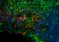
Confocal image of C57BL/6 mouse spleen sample acquired using... 
Confocal image of C57BL/6 mouse thymus sample acquired using... 
Confocal image of C57BL/6 mouse lung sample acquired using t... 
Mice were injected subcutaneously with sheep red blood cells... -
Alexa Fluor® 594 anti-mouse CD68

C57BL/6 mouse frozen spleen section was fixed with 4% parafo... 
Paraformaldehyde-fixed (4%), 500 µm-thick mouse spleen secti... -
APC/Cyanine7 anti-mouse CD68

Thioglycolate-elicited BALB/c macrophages were fixed, permea... -
Brilliant Violet 605™ anti-mouse CD68

Thioglycolate-elicited BALB/c macrophages were fixed, permea... -
Brilliant Violet 711™ anti-mouse CD68

Thioglycolate-elicited BALB/c macrophages were fixed, permea... -
Alexa Fluor® 700 anti-mouse CD68

Thioglycolate-elicited BALB/c macrophages were fixed, permea... -
Pacific Blue™ anti-mouse CD68

Thioglycolate-elicited BALB/c macrophages were fixed, permea... -
TotalSeq™-A0560 anti-mouse CD68
-
TotalSeq™-C0560 anti-mouse CD68
-
Brilliant Violet 785™ anti-mouse CD68

Thioglycolate-elicited Balb/c peritoneal macrophages were su... -
TotalSeq™-B0560 anti-mouse CD68
-
Spark YG™ 570 anti-mouse CD68

C57BL/6 mouse frozen spleen section was fixed with 4% parafo... -
APC/Fire™ 750 anti-mouse CD68

Thioglycollate-elicited BALB/c macrophages were fixed, perm...


Follow Us