 Login / Register


- Clone
- MEC14.7 (See other available formats)
- Regulatory Status
- RUO
- Other Names
- Mucosialin
- Isotype
- Rat IgG2a, κ
- Ave. Rating
- Submit a Review
- Product Citations
- publications
MEC14dot7_Alx647_080607")
-
MEC14dot7_Alx647_080607")
Mouse NIH/3T3 cell line stained with MEC14.7 Alexa Fluor® 647 -

Mouse NIH/3T3 cells were fixed with 1% paraformaldehyde (PFA), and then stained with 1 µg/ml of CD34 (clone MEC14.7) Alexa Fluor® 647 (yellow) for 4 hours at room temperature. Nuclei were counterstained with DAPI and are shown in blue. The image was captured by 40X objective.
| Cat # | Size | Price | Quantity Check Availability | Save | ||
|---|---|---|---|---|---|---|
| 119314 | 100 µg | 193€ | ||||
CD34 is a highly glycosylated hematopoietic progenitor antigen. Two isoforms of CD34 have been reported to be generated by alternative splicing. This antigen is expressed on hematopoietic progenitors as well as on endothelial cells, brain, and testis. CD34 is thought to function as an adhesion molecule for early hematopoietic progenitors mediating the attachment of stem cells to extracellular matrix or stromal cells. CD34 is phosphorylated on serine residues by PKC.
Product DetailsProduct Details
- Verified Reactivity
- Mouse
- Antibody Type
- Monoclonal
- Host Species
- Rat
- Immunogen
- Cells transfected with mouse CD34
- Formulation
- Phosphate-buffered solution, pH 7.2, containing 0.09% sodium azide.
- Preparation
- The antibody was purified by affinity chromatography and conjugated with Alexa Fluor® 647 under optimal conditions.
- Concentration
- 0.5 mg/ml
- Storage & Handling
- The antibody solution should be stored undiluted between 2°C and 8°C, and protected from prolonged exposure to light. Do not freeze.
- Application
-
FC - Quality tested
ICC - Verified - Recommended Usage
-
Each lot of this antibody is quality control tested by immunofluorescent staining with flow cytometric analysis. For flow cytometric staining, the suggested use of this reagent is ≤1.0 µg per million cells in 100 µl volume. It is recommended that the reagent be titrated for optimal performance for each application.
* Alexa Fluor® 647 has a maximum emission of 668 nm when it is excited at 633 nm / 635 nm.
Alexa Fluor® and Pacific Blue™ are trademarks of Life Technologies Corporation.
View full statement regarding label licenses - Excitation Laser
-
Red Laser (633 nm)
- Application Notes
-
The MEC14.7 antibody does not stain bone marrow cells like some other mouse CD34 antibodies, probably because the antibody recognizes a different epitope from other mAbs. Additional reported applications (for the relevant formats) include: immunoprecipitation, Western blotting6, and immunohistochemistry of acetone-fixed frozen sections and paraffin-embedded sections2,4,5,6.
- Application References
-
- Garlanda C, et al. 1997. Eur. J. Cell Biol. 73:368. (FC)
- Knowles HJ, et al. 2004. Circ. Res. 95:162. (IHC)
- Trempus CS, et al. 2003. J. Invest. Dermatol. 120:501.
- Winding B, et al. 2002. Clin. Cancer Res. 8:1932. (IHC)
- Voswinckel R, et al. 2003. Circ. Res. 93:372. (IHC)
- Kairaitis LK, et al. 2005. Am. J. Physiol. Renal. Physiol. 288:F198. (IHC, WB)
- Ao A, et al. 2008. P. Natl. Acad. Sci. USA 105:7821. PubMed
- Zaynagetdinov R., et al. 2011. J Immunol. 187:5703. PubMed.
- Product Citations
-
- RRID
-
AB_604089 (BioLegend Cat. No. 119314)
Antigen Details
- Structure
- Type I membrane protein, 75-120 kD, highly glycosylated; two isoforms reported
- Distribution
-
Hematopoietic progenitors, brain, testis, endothelial cells; low expression in thymus, spleen, and bone marrow
- Function
- Possible adhesion molecule thought to function in early hematopoiesis by mediating attachment of stem cells to bone marrow extracellular matrix or stromal cells. Presents carbohydrate ligands to selectins.
- Ligand/Receptor
- L-selectin, other selectins
- Cell Type
- Endothelial cells, Hematopoietic stem and progenitors
- Biology Area
- Cell Biology, Immunology, Neuroinflammation, Neuroscience
- Molecular Family
- Adhesion Molecules, CD Molecules
- Antigen References
-
1. Garlanda C, et al. 1997. Eur. J. Cell Biol. 73:368.
2. Brown J, et al. 1991. Int. Immunol. 3:175.
3. Suda J, et al. 1992. Blood 79:2288.
4. Baumhueter S, et al. 1994. Blood 84:2554. - Gene ID
- 12490 View all products for this Gene ID
- UniProt
- View information about CD34 on UniProt.org
Related Pages & Pathways
Pathways


Related FAQs
Other Formats
View All CD34 Reagents Request Custom Conjugation| Description | Clone | Applications |
|---|---|---|
| Purified anti-mouse CD34 | MEC14.7 | FC,ICC,IHC-F,IP,WB |
| Biotin anti-mouse CD34 | MEC14.7 | FC,IHC-F |
| PE anti-mouse CD34 | MEC14.7 | FC |
| Alexa Fluor® 647 anti-mouse CD34 | MEC14.7 | FC,ICC |
| PE/Cyanine5 anti-mouse CD34 | MEC14.7 | FC |
| APC anti-mouse CD34 | MEC14.7 | FC |
| Brilliant Violet 421™ anti-mouse CD34 | MEC14.7 | FC,ICC |
| PE/Cyanine7 anti-mouse CD34 | MEC14.7 | FC |
| PerCP/Cyanine5.5 anti-mouse CD34 | MEC14.7 | FC |
| PE/Dazzle™ 594 anti-mouse CD34 | MEC14.7 | FC |
Customers Also Purchased
Compare Data Across All Formats
This data display is provided for general comparisons between formats.
Your actual data may vary due to variations in samples, target cells, instruments and their settings, staining conditions, and other factors.
If you need assistance with selecting the best format contact our expert technical support team.
-
Purified anti-mouse CD34

Mouse NIH/3T3 cell line stained with purified MEC14.7, follo... 
NIH3T3 cells were fixed with 1% PFA and then stained with 10... 
C57BL/6 mouse frozen spleen section was fixed with 4% parafo... -
Biotin anti-mouse CD34

Mouse NIH/3T3 cell line stained with biotinylated MEC14.7, f... 
C57BL/6 mouse frozen spleen section was fixed with 4% parafo... -
PE anti-mouse CD34

Mouse NIH/3T3 cell line stained with MEC14.7 PE -
Alexa Fluor® 647 anti-mouse CD34
MEC14dot7_Alx647_080607")
Mouse NIH/3T3 cell line stained with MEC14.7 Alexa Fluor® 64... 
Mouse NIH/3T3 cells were fixed with 1% paraformaldehyde (PFA... -
PE/Cyanine5 anti-mouse CD34

Mouse NIH/3T3 cell line stained with MEC14.7 PE/Cyanine5 -
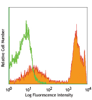
APC anti-mouse CD34

NIH3T3 cells stained with MEC14.7APC -
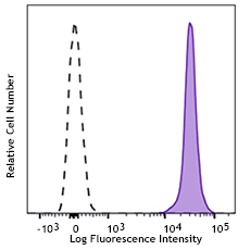
Brilliant Violet 421™ anti-mouse CD34

Mouse fibroblast cell line NIH/3T3 was stained with CD34 (cl... MEC14.7_BV421_041811")
Mouse fibroblast cell line NIH/3T3 was stained with CD34 (cl... -
PE/Cyanine7 anti-mouse CD34

Mouse fibroblast cell line NIH/3T3 was stained with CD34 (c... -
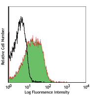
PerCP/Cyanine5.5 anti-mouse CD34

Mouse fibroblast cell line NIH/3T3 was stained with CD34 (cl... -
PE/Dazzle™ 594 anti-mouse CD34

Mouse fibroblast cell line NIH/3T3 was stained with CD34 (cl...


Follow Us