 Login / Register


- Clone
- C-11 (See other available formats)
- Regulatory Status
- RUO
- Other Names
- Cytokeratin, Keratin
- Isotype
- Mouse IgG1, κ
- Ave. Rating
- Submit a Review
- Product Citations
- publications

-

MCF-7 cells were fixed with 1% paraformaldehyde (PFA) for 10 minutes at 37°C, permeabilized with 0.5% TritonX-100 buffer for 10 minutes at room temperature, and blocked for 30 minutes at room temperature with 5% fetal bovine serum (FBS). Then the cells were stained with 5 µg/mL anti-cytokeratin (pan reactive) (clone C-11) Alexa Fluor® 647 (red). Nuclei were counterstained with DAPI (blue). Images were acquired with a 60X objective. -

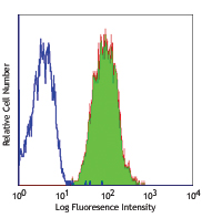
MCF7 cells were fixed and permeabilized using Cyto-Fast Fix/Perm Buffer (Cat. No. 426803) and intracellularly stained with Alexa Fluor® 647 anti-Cytokeratin (Cat. No. 628604) (Filled histogram), or with Alexa Fluor® 647 isotype control (Cat. No. 400135) (Open histogram). -

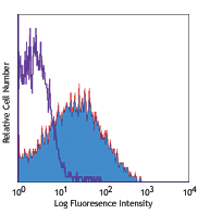
MCF7 cells (positive control, solid histogram), and Jurkat cells (negative control, open histogram) were fixed and permeabilized using Cyto-Fast Fix/Perm Buffer (Cat. No. 426803) and intracellularly stained with Alexa Fluor® 647 anti-Cytokeratin (Cat. No. 628604). -

Confocal image of C57BL/6 mouse thymus sample acquired using the IBEX method of highly multiplexed antibody-based imaging: CD206 (cyan) in Cycle 4 and Cytokeratin (purple) in Cycle 5. Tissues were prepared using ~1% (vol/vol) formaldehyde and a detergent. Following fixation, samples are immersed in 30% (wt/vol) sucrose for cryoprotection. Images are courtesy of Drs. Andrea J. Radtke and Ronald N. Germain of the Center for Advanced Tissue Imaging (CAT-I) in the National Institute of Allergy and Infectious Diseases (NIAID, NIH).
| Cat # | Size | Price | Quantity Check Availability | Save | ||
|---|---|---|---|---|---|---|
| 628604 | 100 µg | 278€ | ||||
Cytokeratins are intermediate filament proteins that are widely expressed in many tissues. These proteins are vital components of the cytoskeleton and interact with a number of other proteins. Cytokeratins are usually found in the cytoplasm,although some cytokeratins can be found in the nucleus and the nucleolus.
Product DetailsProduct Details
- Verified Reactivity
- Human
- Antibody Type
- Monoclonal
- Host Species
- Mouse
- Immunogen
- Keratin enriched fraction from human epidermoid carcinoma.
- Formulation
- Phosphate-buffered solution, pH 7.2, containing 0.09% sodium azide.
- Preparation
- The antibody was purified by affinity chromatography and conjugated with Alexa Fluor® 647 under optimal conditions.
- Concentration
- 0.5 mg/ml
- Storage & Handling
- The antibody solution should be stored undiluted between 2°C and 8°C, and protected from prolonged exposure to light. Do not freeze.
- Application
-
ICC - Quality tested
ICFC - Verified
SB - Reported in the literature, not verified in house - Recommended Usage
-
Each lot of this antibody is quality control tested by immunocytochemistry. For immunocytochemistry, a concentration range of 1.0 - 5.0 μg/ml is recommended. For flow cytometric staining, the suggested use of this reagent is ≤ 0.125 µg per million cells in 100 µL volume. It is recommended that the reagent be titrated for optimal performance for each application.
* Alexa Fluor® 647 has a maximum emission of 668 nm when it is excited at 633 nm / 635 nm.
Alexa Fluor® and Pacific Blue™ are trademarks of Life Technologies Corporation.
View full statement regarding label licenses - Excitation Laser
-
Red Laser (633 nm)
- Application Notes
-
The C-11 monoclonal antibody reacts with a conserved epitope of human cytokeratin 4, 5, 6, 8, 10, 13, and 18. This antibody has been shown to be useful for Western blotting and has also been reported to be useful for immunoprecipitation, immunohistochemistry (paraffin sections), immunocytochemistry, and spatial biology (IBEX)7,8.
- Additional Product Notes
-
Iterative Bleaching Extended multi-pleXity (IBEX) is a fluorescent imaging technique capable of highly-multiplexed spatial analysis. The method relies on cyclical bleaching of panels of fluorescent antibodies in order to image and analyze many markers over multiple cycles of staining, imaging, and, bleaching. It is a community-developed open-access method developed by the Center for Advanced Tissue Imaging (CAT-I) in the National Institute of Allergy and Infectious Diseases (NIAID, NIH).
- Application References
-
- Kovarik J. 1988. Int. J. Cancer Suppl. 3:50.
- Bartek J, et al. 1991. J. Pathol. 164:215.
- Chernyavsky A I, 2007. J. Biol. Chem. doi:10.1074/jbc.M611365200. PubMed
- Comito G, et al. 2012. Cancer Lett. 324:31. PubMed
- Vojt-òsek B, et al. 1989. Folia Biol (Praha). 35:373. (reactivity with rat, dog, sheep, pig, cow)
- Dekaney CM, et al. 2005. Gastroenterology 129:1567. (ICC)
- Radtke AJ, et al. 2020. Proc Natl Acad Sci U S A. 117:33455-65. (SB) PubMed
- Radtke AJ, et al. 2022. Nat Protoc. 17:378-401. (SB) PubMed
- Product Citations
-
- RRID
-
AB_2563652 (BioLegend Cat. No. 628604)
Antigen Details
- Structure
- Intermediate filament protein, contains three coiled-coil domains. Cytokeratins exist as heterotetramers composed of two type I and two type II keratin subunits
- Distribution
-
Cytokeratins are widely expressed proteins usually found in the cytoplasm although some cytokeratins can also be found in the nucleus and the nucleolus
- Function
- Intermediate filament protein involved with the cytoskeleton.
- Interaction
- Most cytokeratins interact with a number of other proteins such as 14-3-3, EGF receptor, and many others
- Biology Area
- Cell Biology, Cell Motility/Cytoskeleton/Structure, Neuroscience, Neuroscience Cell Markers
- Molecular Family
- Intermediate Filaments
- Gene ID
- 3875 View all products for this Gene ID
- UniProt
- View information about Cytokeratin pan reactive on UniProt.org


Related FAQs
- If an antibody clone has been previously successfully used in IBEX in one fluorescent format, will other antibody formats work as well?
-
It’s likely that other fluorophore conjugates to the same antibody clone will also be compatible with IBEX using the same sample fixation procedure. Ultimately a directly conjugated antibody’s utility in fluorescent imaging and IBEX may be specific to the sample and microscope being used in the experiment. Some antibody clone conjugates may perform better than others due to performance differences in non-specific binding, fluorophore brightness, and other biochemical properties unique to that conjugate.
- Will antibodies my lab is already using for fluorescent or chromogenic IHC work in IBEX?
-
Fundamentally, IBEX as a technique that works much in the same way as single antibody panels or single marker IF/IHC. If you’re already successfully using an antibody clone on a sample of interest, it is likely that clone will have utility in IBEX. It is expected some optimization and testing of different antibody fluorophore conjugates will be required to find a suitable format; however, legacy microscopy techniques like chromogenic IHC on fixed or frozen tissue is an excellent place to start looking for useful antibodies.
- Are other fluorophores compatible with IBEX?
-
Over 18 fluorescent formats have been screened for use in IBEX, however, it is likely that other fluorophores are able to be rapidly bleached in IBEX. If a fluorophore format is already suitable for your imaging platform it can be tested for compatibility in IBEX.
- The same antibody works in one tissue type but not another. What is happening?
-
Differences in tissue properties may impact both the ability of an antibody to bind its target specifically and impact the ability of a specific fluorophore conjugate to overcome the background fluorescent signal in a given tissue. Secondary stains, as well as testing multiple fluorescent conjugates of the same clone, may help to troubleshoot challenging targets or tissues. Using a reference control tissue may also give confidence in the specificity of your staining.
- How can I be sure the staining I’m seeing in my tissue is real?
-
In general, best practices for validating an antibody in traditional chromogenic or fluorescent IHC are applicable to IBEX. Please reference the Nature Methods review on antibody based multiplexed imaging for resources on validating antibodies for IBEX.
Other Formats
View All Cytokeratin Reagents Request Custom Conjugation| Description | Clone | Applications |
|---|---|---|
| Purified anti-Cytokeratin (pan reactive) | C-11 | WB,ICC,IP,IHC-P |
| Alexa Fluor® 647 anti-Cytokeratin (pan reactive) | C-11 | ICC,SB,ICFC |
| Alexa Fluor® 594 anti-Cytokeratin (pan reactive) | C-11 | ICC |
| Alexa Fluor® 488 anti-Cytokeratin (pan reactive) | C-11 | ICC,IHC-P |
Customers Also Purchased
Compare Data Across All Formats
This data display is provided for general comparisons between formats.
Your actual data may vary due to variations in samples, target cells, instruments and their settings, staining conditions, and other factors.
If you need assistance with selecting the best format contact our expert technical support team.
-
Purified anti-Cytokeratin (pan reactive)

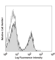
Whole cell extracts (15 µg protein) from Jurkat (negative co... 
MCF-7 cells were stained with purified anti- Cytokeratin (pa... 
Hela cells stained with purified mouse monoclonal antibody a... -
Alexa Fluor® 647 anti-Cytokeratin (pan reactive)

MCF-7 cells were fixed with 1% paraformaldehyde (PFA) for 10... 
Confocal image of C57BL/6 mouse thymus sample acquired using... 
MCF7 cells were fixed and permeabilized using Cyto-Fast Fix/... 
MCF7 cells (positive control, solid histogram), and Jurkat c... -
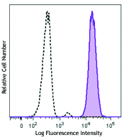
Alexa Fluor® 594 anti-Cytokeratin (pan reactive)

MCF-7 cells were fixed with 1% paraformaldehyde (PFA) for te... -
Alexa Fluor® 488 anti-Cytokeratin (pan reactive)

MCF-7 mammary adenocarcinoma cells were fixed, permeabilized... 
Human paraffin-embedded colon tissue slices were prepared wi...


Follow Us