 Login / Register


- Clone
- BCL/10C4 (See other available formats)
- Regulatory Status
- RUO
- Other Names
- B-cell leukemia 2
- Isotype
- Mouse IgG1, κ
- Ave. Rating
- Submit a Review
- Product Citations
- publications

-

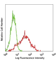
C57BL/6 splenocytes intracellularly stained with BCL/10C4 Alexa Fluor® 647. Cells were fixed and permeabilized with FOXP3 Fix/Perm Buffer Set.
| Cat # | Size | Price | Quantity Check Availability | Save | ||
|---|---|---|---|---|---|---|
| 633509 | 25 µg | 104€ | ||||
| 633510 | 100 µg | 240€ | ||||
Bcl-2 (B-cell leukemia 2) is an apoptotic protein and a member of the Bcl-2 family containing BH1-4 domains. Two reported isoforms exist α=25 kD; β=22 kD. The Bcl-2 protein forms homo- or hetero-dimers with other Bcl-2 family members. Bcl-2 is distributed in the outer mitochondrial membrane, the nuclear envelope, and the endoplasmic reticulum. This protein blocks apoptotic death by controlling mitochondrial membrane permeability. Cleavage of Bcl-2 can convert to pro-apoptotic (by cleavage of BH4 domain). Bcl-2 has been reported to regulate cell cycle progression via ROS. This protein is modified by ASK1/JNK1, PKC, ERKs, and stress-activated kinase phosphorylation and can be ubiquitinated. Bcl-2 has been shown to interact with Apaf-1, Raf-1, TP53BP2, caspase-3, and form heterodimers with Bax, Bad, Bak, Bcl-xL, and Bag-1. Clone BCL/10C4 has been shown to be useful for Western blotting, immunoprecipitation, and immunofluorescence of the mouse and rat Bcl-2 protein.
Product DetailsProduct Details
- Verified Reactivity
- Mouse, Rat
- Antibody Type
- Monoclonal
- Host Species
- Mouse
- Immunogen
- N-terminal, Amino acid residues 61-76 of mouse Bcl-2
- Formulation
- Phosphate-buffered solution, pH 7.2, containing 0.09% sodium azide.
- Preparation
- The antibody was purified by affinity chromatography and conjugated with Alexa Fluor® 647 under optimal conditions.
- Concentration
- 0.5 mg/ml
- Storage & Handling
- The antibody solution should be stored undiluted between 2°C and 8°C, and protected from prolonged exposure to light. Do not freeze.
- Application
-
ICFC - Quality tested
- Recommended Usage
-
Each lot of this antibody is quality control tested by intracellular immunofluorescent staining with flow cytometric analysis. For flow cytometric staining, the suggested use of this reagent is ≤ 1.0 µg per 106 cells in 100 µl volume. It is recommended that the reagent be titrated for optimal performance for each application.
* Alexa Fluor® 647 has a maximum emission of 668 nm when it is excited at 633nm / 635nm.
Alexa Fluor® and Pacific Blue™ are trademarks of Life Technologies Corporation.
View full statement regarding label licenses - Excitation Laser
-
Red Laser (633 nm)
- Application Notes
-
Additional reported applications (for the relevant formats) include: immunohistochemical staining of frozen tissue4, immunocytochemical staining5, and immunoprecipitation5.
- Application References
- Product Citations
-
- RRID
-
AB_2064149 (BioLegend Cat. No. 633509)
AB_2274702 (BioLegend Cat. No. 633510)
Antigen Details
- Structure
- Apoptosis regulator proteins, Bcl-2 family, BH1-4 domains. Homo- or hetero-dimer; isoforms α, β 25 kD, 22 kD
- Distribution
-
Outer mitochondrial membrane, intracellular membrane nuclear envelope, endoplasmic reticulum
- Function
- Blocks apoptotic death by controlling the mitochondrial membrane permeability. Converted to pro-apoptotic activity by cleavage of BH4 domain. Regulates cell cycle progression via ROS
- Modification
- Phosphorylation, Ubiquitination
- Biology Area
- Apoptosis/Tumor Suppressors/Cell Death, Cell Biology, Neuroscience
- Antigen References
-
1. Tsujimoto Y, et al. 1986 P. Natl. Acad. Sci. USA 83:5214.
2. Yang E, et al. 1995. Cell 80:285.
3. Huang Z, et al. 2000. Oncogene 19:6627.
4. Deng X, et al. 2003. Blood. 102:3179. - Regulation
- Phosphorylation by ASK1/JNK1, PKC, ERKs, stress-activated kinases
- Gene ID
- 596 View all products for this Gene ID
- UniProt
- View information about Bcl-2 on UniProt.org
Related Pages & Pathways
Pathways


Related FAQs
Other Formats
View All Bcl-2 Reagents Request Custom Conjugation| Description | Clone | Applications |
|---|---|---|
| Purified anti-Bcl-2 | BCL/10C4 | WB,IP,ICC,IHC-F |
| FITC anti-Bcl-2 | BCL/10C4 | ICFC |
| Alexa Fluor® 488 anti-Bcl-2 | BCL/10C4 | ICFC |
| PE anti-Bcl-2 | BCL/10C4 | ICFC |
| Alexa Fluor® 647 anti-Bcl-2 | BCL/10C4 | ICFC |
| PE/Cyanine7 anti-Bcl-2 | BCL/10C4 | ICFC |
| Direct-Blot™ HRP anti-Bcl-2 | BCL/10C4 | WB |
Customers Also Purchased
Compare Data Across All Formats
This data display is provided for general comparisons between formats.
Your actual data may vary due to variations in samples, target cells, instruments and their settings, staining conditions, and other factors.
If you need assistance with selecting the best format contact our expert technical support team.
-
Purified anti-Bcl-2

Western blot analysis of extracts from mouse splenocytes usi... -
FITC anti-Bcl-2

C57BL/6 splenocytes intracellular stained with BCL/10C4 FITC... -
Alexa Fluor® 488 anti-Bcl-2

C57BL/6 splenocytes intracellular stained with BCL/10C4 Alex... -
PE anti-Bcl-2

C57BL/6 splenocytes intracellularly stained with BCL/10C4 PE... -
Alexa Fluor® 647 anti-Bcl-2

C57BL/6 splenocytes intracellularly stained with BCL/10C4 Al... -
PE/Cyanine7 anti-Bcl-2

C57BL/6 splenocytes were fixed and permeabilized with Nuclea... -
Direct-Blot™ HRP anti-Bcl-2

Western blot analysis of 15 µg cell lysates from Mouse...


Follow Us