Uncovering SARS-CoV-2 Specific Cells and Biomarkers


As we seek to better understand the mechanisms of SARS-CoV-2, it is important to have the right resources on hand to study the corresponding immune response generated by host cells. The first step involves the identification of participating immune cells, like T cells and B cells. This is followed by the thorough characterization and phenotyping of the cells, potentially leading to the discovery of new biomarkers that differentiate subpopulations, effectively opening the way to new treatments and therapies. In this blog post, we will look at new tools BioLegend provides to researchers performing this critical discovery work, including TotalSeq™ proteogenomics antibodies recently used by Su et al. in their Cell publication.


To stay current on our newest SARS-CoV-2 research tools, sign up for our eNewsletter and browse through our webpage for reagents.


TotalSeq™ Reveals Unique Subpopulations


Immune profile changes in moderate/severe COVID-19 patients


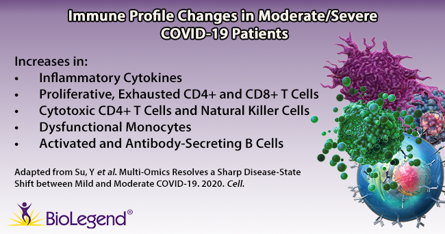
Su et al. began by gathering plasma and PBMC samples from healthy and COVID-19 afflicted patients. Patients were classified as mild, moderate, or severe based on the World Health Organization’s ordinal scale. While the group looked at hundreds of metabolites and secreted proteins, we’ll focus on key immune cell subpopulations they discovered using a panel of over 190 TotalSeq™ oligonucleotide-conjugated antibodies for single-cell multiomic analysis. Interestingly, they found several unique cell subtypes that began to appear in moderate cases, indicating a sharp distinction in the immune cell profile as patients progress from mild to moderate status. Throughout the article, T cell exhaustion is mentioned. T cell exhaustion describes a dysfunctional state where T cells progressively lose cytotoxic or anti-viral function. These cells become exhausted because they are continually exposed to antigens, such as in cancer or chronic viral infections. Phenotypically, these cells may alter their transcriptional and cytokine profile and upregulate exhaustion markers like PD-11. Knowing this, we can review some of the subpopulations identified below.


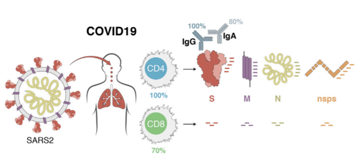
T cells: TCR profiling revealed two sub-populations of CD8+ T cells that correlated with disease severity: cytotoxic effector and memory-like phenotypes. The cytotoxic cells were found mainly in moderate/severe cases, while the memory-like phenotype was from mild patients. This indicates that cells may shift from a memory phenotype to focus on clonal expansion during the progression from a mild to moderate status. In addition, an exhausted and proliferative CD8+ T cell population emerged, as noted by expression of markers like LAG3, TIGIT, and CD279. Within CD4+ T cells, two subpopulations were revealed: one exhibited cytotoxic transcripts for PRF1 and GNLY, while the second featured exhausted and proliferative markers and a Th1 signature phenotype (similar to the CD8+ subpopulation of T cells noted above).


B cells: Patients with moderate/severe cases of COVID-19 possessed B cells that expressed more activation markers and had a larger expansion of antibody-secreting cells compared to healthy/mild cases.


Monocytes: Moderate patients displayed lower levels of non-classical (CD14lowCD16high) monocytes, but an increase in S100high HLA-DRlow monocytes. Su et al. theorize this latter group of monocytes may exhibit dysfunctional tendencies and contribute to a proinflammatory environment.


Natural Killer (NK) cells: NK cells in moderate patients become more cytotoxic-like, exhibiting a decrease in CD56 and CD127 transcripts, but an increase in PRF1, GZMB, and CD69 transcripts.


The shift in disease state from mild to moderate coincides with a rise in these rare cell populations and changes in the metabolic and cytokine profile. Taken together, these results indicate that the moderate stage may serve as the best potential time for therapeutic intervention.


Pinpointing SARS-CoV-2 Antigen Specific T Cells


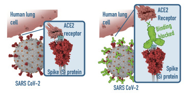
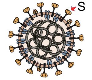
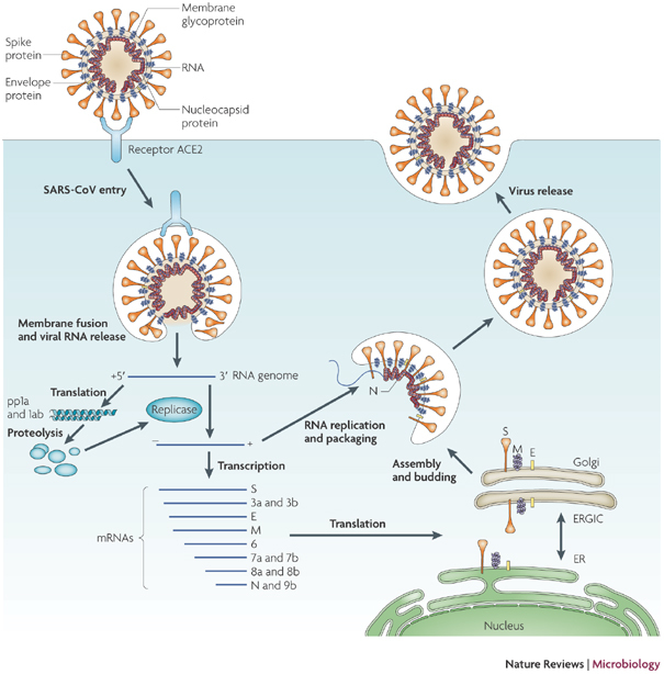
T cells are a key component of the adaptive immune response against SARS-CoV-2. As new therapeutics and vaccine options are developed, such as the latest ones by Pfizer/BioNTech and Moderna, it is paramount to determine which portion of the virus T cells triggers the most efficient response. Our Flex-T™ MHC tetramer technology allows researchers to pinpoint antigen-specific T cells. Provided in monomer form and preloaded with SARS-CoV-2 spike, membrane, or nucleoprotein peptides, these reagents can be assembled into tetramers to stain reacting T cells. A short list of our options can be found below, but dozens more will be released in the coming months. Check our webpage often for updates.


Flex-T™ Peptide SARS-CoV-2 Component

TLACFVLAAV

Membrane protein (61-70)

GMSRIGMEV

Nucleoprotein (316-324)

LLLDRLNQL

Nucleoprotein (222-230)

SIIAYTMSL

Spike protein (691-699)

ALNTLVKQL

Spike protein (958-966)

VLNDILSRL

Spike protein (976-984)

LITGRLQSL

Spike protein (996-1004)

VVFLHVTYV

Spike protein (1042-1050)

RLNEVAKNL

Spike protein (1185-1193)

NLNESLIDL

Spike protein (1192-1200)

FIAGLIAIV

Spike protein (1220-1228)


Additional Reagents for SARS-CoV-2 Research


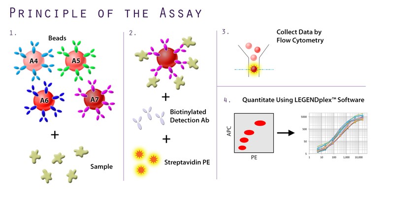
In addition to the unique offerings above, we provide several immunoassays for your SARS-CoV-2 workflow, including: MojoSort™ for magnetic bead-based cell isolation; recombinant proteins, cytokines, and growth factors for bioassays; and LEGENDScreen™, which allows for the screening of hundreds of cell surface markers via flow cytometry. Although the methodology differs between the immunoassays, both LEGENDScreen™ and TotalSeq™-C Human Universal Cocktail, V1.0 have the ability to detect a variety of surface proteins. The latter reagent was utilized in the paper by Su et al. and is  available as a ready-to-use cocktail with 130 markers and 7 isotype control antibodies.


In order for research to progress, a versatile set of tools must be made available to researchers, clinicians, and scientists. BioLegend will continue to develop these novel reagents so that we can fully understand each cell’s role in SARS-CoV-2 immunity.


T cells – the Unsung Heroes of the Anti-SARS-CoV-2 Immune Response?


T cells are critical for immunity against viruses and understanding their response to SARS-CoV-2 will be critical for scientists to design effective vaccines. However, recent attention has focused on antibodies – the other arm of the adaptive immune response. This week, we will shine a spotlight on how researchers are using a diverse set of computational, immunological, and single-cell tools to decipher the role of SARS-CoV-2 specific T cells. Could T cells be the key to defeating COVID-19?


Bioinformatics Predict Targets for SARS-CoV-2-Specific T Cells


Early in the pandemic when data on the immune response to SARS-CoV-2 was scarce, researchers relied on bioinformatics to predict potential SARS-CoV-2 T cell epitopes – which are short viral peptides recognized by the T cell receptor.


A study in Cell Host and Microbe used two independent computational methods to find SARS-CoV-2 peptides with a high likelihood of being targeted by T cells. First, the authors mined existing data for original SARS-CoV peptides known to be recognized by T cells from SARS patients. Alignment of these peptides’ sequences to the SARS-CoV-2 genome identified their SARS-CoV-2 equivalents or best matches. For the second approach, the authors used algorithms to scan the SARS-CoV-2 genome and predict peptide sequences with the highest potential as T cell targets. In the end, both methods – sequence alignment and algorithm prediction – led to the identification of the same peptides. These peptides are now synthesized and used for various research applications.


Viral peptides are commonly used to identify virus-specific T cells through tetramer staining. Tetramers loaded with SARS-CoV-2 peptides can bind to T cell receptors that recognize those peptides. Since the tetramers are labeled with a fluorescent marker, they allow researchers to detect virus-specific T cells by flow cytometry.


BioLegend Flex-T™ reagents, which come pre-loaded with SARS-CoV-2 peptides discovered in the study mentioned above, can streamline your workflow for virus-specific T cell analysis. Browse SARS-CoV-2 Flex-T™ reagents here or learn more about how Flex-T™ works.


Multiplex Immunoassays Classify the T Cell Response in COVID-19 Patients


In addition to tetramer staining, peptides can also be used to stimulate virus-specific T cells. When T cells with immunological memory of SARS-CoV-2 are presented again with the same viral peptides, they will express specific activation markers. Two studies published in Cell and Science Immunology examined the blood of SARS-CoV-2-infected individuals, and they looked for T cells expressing these activation markers following SARS-CoV-2 peptide stimulation. Out of the combined 30 people analyzed, almost all of them had SARS-CoV-2 peptide-reactive CD4+ and CD8+ T cells. Disease in the patients studied ranged from mild to severe, suggesting most people infected with SARS-CoV-2 – even those with light symptoms – generate a virus-specific T cell response to some degree.


Though activation markers reveal the presence of virus-specific T cells, these markers don’t tell us how the cells respond. One way to characterize the functional response of peptide-stimulated CD4+ T helper cells is to measure their cytokine production. In both of these studies, the researchers utilized BioLegend’s LEGENDplex™ T Helper Cytokine Panel to simultaneously measure 12 cytokines released by T cells from patient blood. They discovered that the cytokines produced by virus-specific CD4+ T cells were predominantly of the Th1 type – such as IFN-γ and IL-2. Though cytokines of other T helper types were also detected, they were produced at relatively low levels and only observed in T cells from patients with more severe disease.


Grifoni et al. discovered the presence of T cells specific for multiple SARS-CoV-2 proteins in 70-100% of infected individuals. Image source: Grifoni et al. 2020 Cell, DOI: https://doi.org/10.1016/j.cell.2020.05.015


Using these two “read-outs” – activation markers and cytokines – the researchers made two more notable discoveries about SARS-CoV-2-specific T cells.


First, T cells from COVID-19 patients recognized multiple SARS-CoV-2 proteins, and they did not show a strong preference for the spike as they do for other coronaviral infections. The researchers detected T cells reactive to at least 6 out of the 25 proteins encoded by SARS-CoV-2 in patient blood. Based on this data, the authors concluded that vaccines eliciting T cells against multiple viral proteins may be more effective than vaccines eliciting T cells against just a single target.


Second, up to 60% of the SARS-CoV-2-negative people analyzed also had SARS-CoV-2-specific T cells. This suggests that people who have been previously infected with other coronaviruses – such as ones causing common colds – can develop cross-reactive T cells against SARS-CoV-2. Though unproven, the researchers speculate that these cross-reactive T cells may offer some level of protection against SARS-CoV-2. In support of this hypothesis, influenza studies have shown T cells responding to the seasonal flu could cross-react with the pandemic H1N1 strain, and people who had these cross-reactive T cells suffered less severe H1N1 disease. More research is needed to know whether cross-reactive T cells are protective against COVID-19.


Learn more about how you can characterize T cell responses, cytokine storm, or other immune markers during SARS-CoV-2 infection with our LEGENDplex™ multiplex immunoassays.


Single-Cell Sequencing Finds Expanded CD8+ T Cells in SARS-CoV-2-Infected Lungs


Another approach for evaluating the T cell response to SARS-CoV-2 is to phenotype individual cells from patient samples. A study published in Nature Medicine used single-cell transcriptome sequencing to characterize lung T cells from nine COVID-19 patients. By looking at the T cell receptor gene transcripts in single T cells, the researchers tracked how many T cells were derived from a single clone in each patient. Severe COVID-19 was associated with less clonal expansion and more heterogeneity of CD8+ T cells when compared with moderate cases. Though the sample size was small, the authors suggest their observations help us understand T cell phenotypes associated with recovery from COVID-19.


Future single-cell studies will help to pinpoint protective T cell clones, and the exact viral peptides they recognize. This would better inform the design of effective vaccines, especially since recent research has shown correlations between SARS-CoV-2-specific T cell numbers and neutralizing antibody titers.


Interested in single-cell multiomics? Improve single-cell phenotyping with BioLegend’s TotalSeq™ reagents, which capture both proteomic and transcriptomic data.


References

  1. Grifoni A et al. A Sequence Homology and Bioinformatic Approach Can Predict Candidate Targets for Immune Responses to SARS-CoV-2. Cell Host and Microbe (2020). DOI: 10.1016/j.chom.2020.03.002
  2. Grifoni A et al. Targets of T Cell Responses to SARS-CoV-2 Coronavirus in Humans with COVID-19 Disease and Unexposed Individuals. Cell (2020). DOI: 10.1016/j.cell.2020.05.015
  3. Weiskopf D et al. Phenotype and kinetics of SARS-CoV-2–specific T cells in COVID-19 patients with acute respiratory distress syndrome. Science Immunology (2020). DOI: 10.1126/sciimmunol.abd2071
  4. Liao M et al. Single-cell landscape of bronchoalveolar immune cells in patients with COVID-19. Nature Medicine (2020). DOI: 10.1038/s41591-020-0901-9
  5. Ni L et al. Detection of SARS-CoV-2-Specific Humoral and Cellular Immunity in COVID-19 Convalescent Individuals. Immunity (2020). DOI: 10.1016/j.immuni.2020.04.023


Antibodies Reject Viral Entry by Exploiting the SARS-CoV-2 Spike Protein


One of the key strengths of SARS-CoV-2 is its spike (S) protein, which enables the virus to bind tightly to its cellular receptor, ACE2. As scientists gain a better understanding of the spike’s characteristics, however, the spike may soon become the virus’ weakness. This week, we discuss new research on antibodies that are leading the attack on the viral spike protein.


Anti-RBD Antibodies Block Spike Binding and Fusion


The spike protein of SARS-CoV-2 has two functional components, S1 and S2. A region on S1 that is extremely valuable to the virus is the receptor-binding domain (RBD), since it is responsible for engaging the spike with ACE2. This initial interaction is what allows the virus to attach to the cell and eventually gain entry. Because RBD binding is the first step in viral infection, it is thought that immunotherapies aimed at neutralizing the virus should target the RBD.


Scientists are now trying to define the exact sections of the RBD that are required for ACE2 binding. Antibodies that target these specific regions within the RBD would likely be the most effective at blocking the spike-ACE2 interaction. To find the precise points of contact, researchers at the Fred Hutchinson Cancer Research Center systematically mutated single amino acids within the RBD sequence and then tested the ability of each mutant to bind to ACE2 [1]. They identified the mutations that had a detrimental impact on binding, which allowed them to map the RBD sections indispensable for ACE2 attachment. The researchers suggest that these particular sections within the RBD are prime targets for immunotherapies, since they are absolutely required for cellular binding.


BioLegend recombinant SARS-CoV-2 S Protein RBD-Fc Chimera binding to recombinant human ACE2. When recombinant SARS-CoV-2 S Protein RBD-Fc Chimera is immobilized at 2 μg/mL, recombinant human ACE2 (Cat. No. 792002) binds in a dose-dependent manner. The EC50 range for this effect is 6 - 30 ng/mL. HRP anti-His Tag antibody (Cat. No. 652503) was used to detect the binding.


Other research suggests that preventing RBD binding to ACE2 isn’t the only way to block infection. Anti-RBD antibodies may also block the second step of viral entry: membrane fusion. A study in Cell Host and Microbe reports that a previously identified antibody against the original SARS-CoV RBD cross-reacts with the SARS-CoV-2 RBD, and can block infection of SARS-CoV-2 in cell culture [2]. The mechanism by which this antibody neutralizes the virus, however, is not through physical obstruction of ACE2 binding. Rather, this antibody triggers conformational changes of the spike. Using cryogenic electron microscopy, the scientists show that binding with this particular antibody alters the structure of the spike protein to a fusion-incompetent shape. This prevents the virus from fusing its membrane with the cellular membrane, and stops the virus from entering the cell even if it docks onto ACE2.


A Multipronged Approach to Neutralization


Even as scientists begin to identify which anti-RBD antibody has the most potential as a drug candidate, it is clear that a single anti-RBD antibody may not be enough to stop SARS-CoV-2. The virus can undergo natural mutations, which form new viral variants or strains that escape recognition by individual antibodies. This raises the need for a multipronged approach to neutralizing the virus.


A recent study published in Science found that growth of SARS-CoV-2 in cell culture under the selective pressure of single anti-RBD antibodies led to the rapid emergence of escape mutants [3]. These mutants were protected from neutralization because their RBD sequences had changed such that they were no longer recognizable by the individual antibodies. However, when the virus was grown in the presence of a cocktail of antibodies targeting unique sites on the RBD, escape mutants were not generated. The researchers conclude that attacking multiple areas of the RBD simultaneously with different antibodies can prevent viral escape. This antibody cocktail is currently being tested in clinical trials.


Inducing Natural Antibody Responses Against the Spike


Development of antibody therapies targeting the spike, though important, is not the ideal end goal. To fully stop the spread of SARS-CoV-2, a vaccine is needed. New research suggests that our immune system, if stimulated correctly, is capable of generating anti-RBD antibodies that neutralize the virus. This bodes well for vaccine candidates using the spike protein to elicit immune responses.


Anti-RBD antibodies blocking spike binding to ACE2. Image source: Suthar et al. Cell Reports Medicine 2020
DOI:
https://doi.org/10.1016/j.xcrm.2020.100040


An analysis of 44 hospitalized COVID-19 patients at Emory University found that anti-RBD antibodies were detectable in the blood of >80% of patients studied [4]. Lack of anti-RBD antibodies in several individuals may have been due to the early timepoints at which their blood was examined – when antibody responses may not yet have peaked. When the researchers tested antibody-containing plasma from these patients in virus-neutralization assays, they found that neutralization ability correlated with anti-RBD antibody concentrations. Their findings indicate that anti-RBD antibodies may be a strong predictor of protective immunity.


Ultimately, vaccines that equip our immune system with the right antibodies will offer the most protection. More knowledge on the spike protein and its points of vulnerability will help scientists achieve this. Research on the spike will also prepare us for a constantly adapting coronavirus family. As new coronaviral spikes evolve, so should our weapons that target them.


BioLegend now offers recombinant SARS-CoV-2 spike proteins and anti-spike antibodies to aid in vaccine, antibody, and viral research. Find them here.


References

  1. Starr T et al. Deep mutational scanning of SARS-CoV-2 receptor binding domain reveals constraints on folding and ACE2 binding. bioRxiv (2020). DOI: 10.1101/2020.06.17.157982
  2. Huo J et al. Neutralization of SARS-CoV-2 by Destruction of the Prefusion Spike. Cell Host and Microbe (2020). DOI: 10.1016/j.chom.2020.06.010
  3. Baum A et al. Antibody cocktail to SARS-CoV-2 spike protein prevents rapid mutational escape seen with individual antibodies. Science (2020). DOI: 10.1126/science.abd0831
  4. Suthar M et al. Rapid Generation of Neutralizing Antibody Responses in COVID-19 Patients. Cell Reports Medicine (2020). DOI: 10.1016/j.xcrm.2020.100040


ACE2: Gateway or Guardian?


Angiotensin converting enzyme 2 (ACE2) is the receptor used by SARS-CoV-2 to gain entry into cells. This week, we discuss a new study that maps the expression of ACE2 in the respiratory tract and the use of recombinant ACE2 proteins to neutralize virus particles.


ACE2 Expression is Associated with Infectivity


A new study published in Cell utilized in situ RNA hybridization to map ACE2 expression along the length of the respiratory tract. Staining of healthy lung epithelial tissue samples with RNA probes showed that ACE2 expression is the highest in the upper respiratory tract (nose and oropharynx) and progressively decreases as the airway reaches the alveoli. To investigate how this expression gradient relates to infectivity by SARS-CoV-2, the researchers developed genetically engineered SARS-CoV-2 reporter viruses that fluoresce after successfully infecting a cell. The researchers found that these reporter viruses replicated to higher levels in upper airway cell cultures compared with lower airway cell cultures, which reflects ACE2 expression patterns. Post-mortem lung tissues from COVID-19 patients also showed signs of this infection gradient [1].


Given these results, the researchers suggested a mechanism for how SARS-CoV-2 travels to the lower respiratory tract. Since lower airway cells express low levels of ACE2 and are difficult to infect directly in culture, accumulation of SARS-CoV-2 in the upper airway may be needed before the virus can achieve infection of the lower tract. When the virus enters through the nose, it is first exposed to cells that have high expression of ACE2. Due to the abundance of receptors, the virus could easily replicate and establish infection in this upper region of the airway before moving down to the lower tract with a much higher concentration of virus. If SARS-CoV-2 infection is seeded in the nose, the researchers speculate that therapies aimed at reducing nasal viral titers could help prevent development of severe disease.

ACE2 expression and infectivity gradient along the respiratory tract. Image source: Hou Y et al. (2020) Cell. DOI: https://doi.org/10.1016/j.cell.2020.05.042


Recombinant ACE2 Therapies


Though membrane-bound ACE2 allows entry of the virus into cells, soluble ACE2 proteins can act as decoy receptors for SARS-CoV-2 and prevent the virus from binding cellular receptors. Recombinant ACE2 proteins have been shown to neutralize the virus in in vitro experiments, demonstrating their therapeutic potential. One limitation of recombinant ACE2 as a drug, however, is its short half-life in vivo. Mouse studies have indicated that recombinant ACE2 proteins are quickly cleared from the body [2].


To increase the half-life of recombinant ACE2 proteins, the authors of a new study published in Nature Communications fused ACE2 to the Fc region of human IgG antibody – an alteration known to lengthen the circulation time of soluble proteins [3]. They called this fusion protein ACE2-Ig. In virus neutralizing assays, ACE2-Ig was able to prevent infection of cells by viruses expressing either the SARS-CoV-2 or SARS-CoV spike proteins. Pharmacokinetic analysis in mice demonstrated that ACE2-Ig had a longer half-life than what has been previously shown for recombinant ACE2 alone. Thus, the researchers propose ACE2-Ig as an alternative approach to recombinant ACE2-mediated blockade of SARS-CoV-2 infection.


Enzymatic activity of BioLegend recombinant human ACE2, as measured by hydrolysis of a fluorogenic substrate.

In addition to preventing SARS-CoV-2 binding and entry into cells, recombinant ACE2 may also have direct anti-inflammatory effects. This is due to ACE2’s physiological function as a regulator of the renin-angiotensin system (RAS). A major component of RAS is angiotensin II, a hormone that signals for blood vessel constriction to increase blood pressure and can cause inflammation in certain experimental models. Angiotensin II is processed by ACE2’s extracellular catalytic domain into angiotensin1-7, a molecule that induces blood vessel dilation and can inhibit inflammation [4].


ACE2 therefore maintains the critical balance between these two opposing molecules, and its enzymatic activity could actually dampen inflammation. Given the potential anti-inflammatory benefits of ACE2, scientists have suggested that treatment of COVID-19 patients with soluble recombinant ACE2 may do more than neutralize the virus: it could also help in limiting inflammatory disease in specific contexts [4]. Since inflammatory cytokines like IL-6 and TNF-α are major drivers of COVID-19, inhibition of inflammatory pathways by recombinant ACE2 would be a welcome side effect.  


If you’re researching the ability of recombinant ACE2 to neutralize SARS-CoV-2 or inhibit inflammation, check out our recombinant human and mouse ACE2 proteins.


References

  1. Hou Y et al. SARS-CoV-2 Reverse Genetics Reveals a Variable Infection Gradient in the Respiratory Tract. Cell (2020). DOI: 10.1016/j.cell.2020.05.042
  2. Wysocki J et al. Targeting the degradation of angiotensin II with recombinant angiotensin-converting enzyme 2: prevention of angiotensin II-dependent hypertension. Hypertension (2010). DOI: 10.1161/HYPERTENSIONAHA.109.138420
  3. Lei C et al. Neutralization of SARS-CoV-2 spike pseudotyped virus by recombinant ACE2-Ig. Nature Communications (2020). DOI: 10.1038/s41467-020-16048-4
  4. Verdecchia P et al. The pivotal link between ACE2 deficiency and SARS-CoV-2 infection. Eur J Intern Med (2020). DOI: 10.1016%2Fj.ejim.2020.04.037


Interferons During SARS-CoV-2 Infection


The interferon (IFN) response is our immune system’s first line of defense against viral infections. As a critical part of innate immunity, IFNs are produced when cells detect the presence of a virus and signal for cells to create an “antiviral state.” This occurs by the activation of interferon-stimulated genes (ISGs), many of which have direct antiviral functions like blocking viral entry or inhibiting viral genome replication. However, coronaviruses are known to subvert the IFN response. This week, we discuss research that uses transcriptome and single-cell analysis to explore the weakened IFN response in COVID-19 patients and the effects of IFN-based treatments.


Impaired IFN Responses to SARS-CoV-2


Evidence is mounting that the IFN response to SARS-CoV-2 infection is impaired. A recent study examined the immune response in the blood of COVID-19 patients at 8-12 days after onset of symptoms [1]. Using RNA sequencing, the researchers found that in patients with severe disease, expression of ISGs was lower in comparison with patients with mild infection. The researchers also found lower levels of blood IFN protein in severe cases than in mild cases, suggesting that a diminished innate antiviral response to SARS-CoV-2 contributes to disease progression. In support of this, they showed that lower IFN levels were associated with higher plasma viral loads and higher concentrations of disease-driving cytokines like IL-6 and TNF-α.


Another study examined the host transcriptional response to SARS-CoV-2 versus influenza virus [2]. Using ferrets as a disease model, the researchers found that SARS-CoV-2 elicited a significantly weaker IFN response than influenza in these animals. To see if their findings in ferrets were relevant to COVID-19 patients, the researchers then measured blood IFN levels in a cohort of hospitalized patients who tested positive for SARS-CoV-2. They found that hospitalized COVID-19 patients had undetectable levels of IFN proteins, while still exhibiting high inflammatory signatures. Taken together, these data suggest that a weakened IFN response to SARS-CoV-2 allows the virus to cause severe disease. But why do our early antiviral defenses fail against this virus?


A Family of Escape Artists


Coronaviruses are known to use a number of strategies to evade the innate antiviral response. The original SARS-CoV is able to avoid detection by pathogen recognition receptors (PRRs) either by shielding its viral RNA or by disrupting PRR activation. Other coronaviruses use their nonstructural proteins to disrupt signaling by interferon regulatory factors (IRFs) – transcriptional regulators of IFN expression. Even after IFNs are produced and start inducing expression of ISGs, coronaviruses have methods to stop these antiviral proteins. One example is the MERS-CoV NS4b protein. This protein is used by the virus to block RNaseL, an IFN-activated nuclease that destroys viral RNA [3].


Methods used by coronaviruses to evade the IFN response.
Image source: Vabret N et al. (2020) Immunity, DOI: https://doi.org/10.1016/j.immuni.2020.05.002
Graphic artist: J. Gregory, Mt. Sinai Health System.


Though the exact anti-IFN mechanisms of SARS-CoV-2 are still unclear, a few studies have revealed protein interactions that indicate how SARS-CoV-2 dampens the innate antiviral response. The SARS-CoV-2 proteins ORF9b and NSP15 have been shown to interact with RIG-I/MAVS and IRF3 signaling, respectively [4]. RIG-I/MAVS and IRF3 are important for triggering the IFN response, so ORF9b and NSP15 likely exert their anti-IFN effects by shutting off these pathways. Thus, SARS-CoV-2 should be no exception in the coronavirus family, a group of viruses that have evolved to be highly adept at escaping innate immunity. These evasion tactics may at least partially explain why some COVID-19 patients develop weak IFN responses to infection.


IFN: A Double-Edged Sword


If the IFN response to SARS-CoV-2 is too weak, the therapeutic answer to this seems straightforward: give patients more IFN. New research cautions that this strategy may not be so simple. A recent study on ACE2, the receptor SARS-CoV-2 uses to gain entry into cells, suggests IFN intervention could actually help the virus replicate. Using single-cell RNA sequencing data, Ziegler et al. characterized human and animal cells expressing ACE2 and found that ACE2 mRNA was often co-expressed with ISGs in the same cell types [5]. This led the researchers to hypothesize that ACE2 expression was also controlled by IFN signaling. Indeed, treatment of human airway epithelial cell cultures resulted in ACE2 mRNA and protein upregulation, indicating that ACE2 itself is an ISG. This means that treating COVID-19 patients with IFNs has the potential to increase infection by stimulating higher expression of the virus’ receptor.


IFN stimulation may upregulate ACE2 expression and increase SARS-CoV-2 infection.
Image source: Ziegler et al 2020 Cell, DOI: https://doi.org/10.1016/j.cell.2020.04.035


IFN-based treatments may therefore be a double-edged sword. Though anecdotal evidence suggests IFN therapies could worsen SARS disease [6], a more precise application could be effective. Timing of IFN intervention may be a key determinant of its success, since studies in mice have shown that early, but not late, IFN treatment could protect animals from coronaviral disease [7, 8]. Additionally, results from a recent phase II clinical trial demonstrated the effectiveness of IFN, if it was combined with the antiviral drug lopinavir, in shortening recovery time from SARS-CoV-2 infection [9]. Perhaps the added benefit of lopinavir’s direct viral inhibition is enough to overcome IFN’s off-target effects.


As the number of potential COVID-19 therapies under investigation continues to grow, learning more about the virus and its disease will help scientists refine our approaches to treatment. Understanding why therapies work – or situations in which they may fail – starts with knowing the basic science.


If you’re studying innate and adaptive immunity against SARS-CoV-2, learn more about the TotalSeq™ workflow and how multimodal techniques for cellular phenotyping can help you characterize immune responses to viral infections.


References

  1. Hadjadj J et al. Impaired type I interferon activity and exacerbated inflammatory responses in severe Covid-19 patients. bioRxiv (2020). DOI: 10.1101/2020.04.19.20068015
  2. Blanco-Melo D et al. Imbalanced host response to SARS-CoV-2 drives development of COVID-19. Cell (2020). DOI: 10.1016/j.cell.2020.04.026
  3. Vabret N et al. Immunology of COVID-19: current state of the science. Immunity (2020). DOI: 10.1016/j.immuni.2020.05.002
  4. Gordon DE et al. A SARS-CoV-2 protein interaction map reveals targets for drug prepurposing. Nature (2020). DOI: 10.1038/s41586-020-2286-9
  5. Ziegler C et al.  SARS-CoV-2 receptor ACE2 is an interferon-stimulated gene in human airway epithelial cells and is detected in specific cell subsets across tissues. Cell (2020). DOI: 10.1016/j.cell.2020.04.035
  6. Lei J et al. CT Imaging of the 2019 Novel Coronavirus (2019-nCoV) Pneumonia. Radiology (2020). DOI: 10.1148/radiol.2020200236
  7. Kindler E et al. SARS-CoV and IFN: Too Little, Too Late. Cell Host and Microbe (2016). DOI: 10.1016%2Fj.chom.2016.01.012
  8. Channappanavar R et al. Dysregulated Type I Interferon and Inflammatory Monocyte-Macrophage Responses Cause Lethal Pneumonia in SARS-CoV-Infected Mice. Cell Host and Microbe (2020). DOI: 10.1016%2Fj.chom.2016.01.007
  9. Hung I et al. Triple combination of interferon beta-1b, lopinavir–ritonavir, and ribavirin in the treatment of patients admitted to hospital with COVID-19: an open-label, randomised, phase 2 trial. The Lancet (2020). DOI: 10.1016/S0140-6736(20)31042-4


LEGENDplex™ Immunoassays for COVID-19 Research


Jason Lehmann

Jason Lehmann, Ph.D., Emerging Technologies Scientist.

Multiplexed cytokine analysis is needed now more than ever as scientists race to understand immune responses to SARS-CoV-2. LEGENDplex™ bead-based immunoassays enable the measurement of multiple soluble analytes in biological samples with flow cytometry – view our video to learn the basic principles of the assay. In this blog post, we ask our LEGENDplex™ expert, Dr. Jason Lehmann, about how these tools can be integrated into your COVID-19 research.


What sample types are appropriate for the LEGENDplex™ assay?


The LEGENDplex™ assay is validated in-house for serum and culture supernatant sample types. We also know that plasma and tissue homogenates can be used with a few caveats. Here are our recommendations for using plasma:


  • Plasma should be collected using an anti-coagulant (e.g., EDTA, Heparin, Citrate) and centrifuged within 30 minutes of blood collection.
  • If the assay can’t be run immediately, aliquot and store samples at ≤-20°C.
  • Avoid multiple (>2) freeze/thaw cycles and when using frozen samples, it is recommended that samples be thawed completely, mixed well and centrifuged to remove particulates.


The use of tissue homogenate samples with the LEGENDplex™ assay has been published in the literature, but has not been validated by BioLegend directly due to the large variety of potential sample inputs. Our general recommendations for using tissue homogenates include:


  • Preparing samples in a neutral pH buffer containing no denaturing chemicals such as SDS and no ionic detergents.
  • Minimal amounts of non-ionic detergents may be used in sample preparation.
  • The tissue/cell homogenization buffer should not contain excessive ionic strength above physiological concentrations of salts.
  • If possible, the buffer should also contain protease inhibitors and samples should be centrifuged to remove particulates.
  • Additionally, we recommend total protein content of each well to be standardized with an amount empirically determined by the investigator.


LEGENDplex™ sounds like it would fit my new COVID-19 research project using human cell supernatants. My samples are from infectious patients. Are there any recommendations on using these samples with the LEGENDplex™ assay?


We have not used COVID-19 patient samples in-house, but we would suggest handling them in the same manner as other infectious samples by fixing them with 1-4% PFA after the completion of the bench portion of the LEGENDplex™ assay and right before analysis on a flow cytometer. This is to limit the amount of time the beads are in contact with fixative solutions.


Now for choosing the right panel for my project. I am interested in analysis of 9 analytes, and I have heard about BioLegend's pre-defined LEGENDplex™ panels, which typically have 13 targets. Do I have the option of creating one that consists of only my analytes of interest?


Yes, you do. We offer three options for picking the LEGENDplex™ panel that suits your project:


  1. First, we offer pre-defined panels that come as a single catalog number such as the Anti-Virus Response Panel.
  2. Second, you can narrow down any of our pre-defined panels into a mix & match system that only contains your analytes of interest. You can purchase each part of the assay as a separate item.
  3. Or third, you can create a whole new combination of analytes in a custom panel with our team’s guidance.

I would be interested in a mix & match that includes cytokine storm-associated signals, such as IL-6, TNF-α, IL-8, IFN-β, and IL-10. When I conduct my assays, should I select the V-bottom or filter plate option?


We recommend a filter plate if you have a vacuum manifold. If not, a V-bottom plate or low-binding polypropylene micro-FACS tubes can be used. Polystyrene ELISA or cell culture plates should not be used. Also, if you do choose a filter plate, be sure to use vacuum pressures that are within the product manual’s specifications.

coronavirus in lungs

Vacuum manifold with filter plate.


Now that I have chosen a panel, are there any resources for seeing the assay performed?


Legendplex Protocol Video Demo

View the LEGENDplex™ protocol demo video.

There are. First, each pre-designed panel comes with a very thorough product manual that can be found on the technical data sheet webpage for each product. We also have a demonstration video that can take you through the entire assay. We encourage customers to be especially mindful of their pipetting when making the standard curve in order to generate the best data possible.


Another particularly important aspect of running the assay is flow cytometer machine settings. We also provide set-up guides for a number of different flow cytometers to ensure sufficient bead separation and accurate signal in the reporter channel. To learn more about our instrument set-up guides, please contact our technical support team.


Once I have run the assay, and I have my FCS files, do I need a special software to analyze it?


Yes, you do. BioLegend offers a cloud-based LEGENDplex™ analysis software that you can use from any computer once you have an active username and password. You can get set-up with an account by contacting our technical support team. I will mention the password set-up email is automated and can sometimes get filtered, so be sure to check your junk folder. View our video tutorial on how to use the software to get started.


If you have further questions about how LEGENDplex™ can help your research, please do not hesitate to reach out to us.


Three Key Stages in the Coronavirus Life Cycle - and Efforts to Combat the Virus


SARS-CoV-2 belongs to the Coronaviridae (or simply, coronavirus) family, a group of single-stranded positive-sense RNA viruses named for their crown-like spikes on the viral surface. Though there may be unique features of SARS-CoV-2 replication that are still unknown, it is believed that SARS-CoV-2 follows the same general life cycle of coronaviruses. This week, we review three key steps in coronavirus replication and discuss recent research on ways to stop each step.


1. Virus entry


coronavirus in lungs

The SARS-CoV-2 spike (S) protein. Image from Virology Blog by Vincent Racaniello.

A coronavirus particle is comprised of an outer membrane envelope, and an internal core built by the virus’ nucleocapsid (or N) proteins. Embedded in the envelope are three structural proteins: the spike (S) protein needed for cellular entry, the membrane (M) protein that gives the virus its curvature, and the envelope (E) protein that allows for release of a newly assembled virus particle from the host cell [1].


S proteins are structured as homotrimers, meaning three identical S protein parts combine to form the functional unit. Each S trimer has two domains important for its function, one domain for receptor binding and one domain for triggering fusion of the viral envelope with the cellular membrane [2]. Both domains are critical to complete the two-step process required for the virus to enter a host cell.


The first entry step relies on the receptor-binding domain (RBD), and the RBDs of both SARS-CoV-2 and the original SARS-CoV (discovered in 2003) recognize angiotensin converting enzyme 2 (ACE2) expressed on human cells [3]. Research on the binding affinity between S proteins and ACE2 found that SARS-CoV-2’s S protein actually binds more tightly to ACE2 than its SARS-CoV counterpart [4]. This may be one reason why SARS-CoV-2 has been so successful as a pathogen.


Following ACE2 binding, the second step required for viral entry involves the cleavage of the S protein’s fusion domain, which possesses a sequence recognized by host cell proteases. Several proteases in human cells are able to complete this action, including cathepsins that cleave cysteines and TMPRSS2 that cleaves serines [5]. Analysis of ACE2 and TMPRSS2 mRNA expression revealed that they can be co-expressed in the same cells across many tissue types [6], indicating that both host factors needed by the virus for entry can be found throughout the body. This is likely what allows SARS-CoV-2 to cause disease in multiple organs, including the nervous system.


Approaches to blocking SARS-CoV-2 entry into cells have focused on targeting these two entry steps: receptor binding and S protein cleavage. Preventing receptor binding relies on disrupting the interaction between the S protein and ACE2, and there are several different ways to do this.


One strategy is to use antibodies. Anti-ACE2 antibodies have been shown to inhibit viral entry in cell cultures [5], and researchers have begun to characterize B cell clones that produce antibodies blocking the S protein’s receptor binding domain [7]. Both antibody-based methods may be viable options for obstructing the S protein-ACE2 interface.


The ability of recombinant ACE2 proteins to “distract” SARS-CoV-2 is also being tested. Recent research showed that soluble ACE2 inhibits SARS-CoV-2 infection of human organoids [8], indicating that decoy ACE2 proteins could potentially outcompete actual ACE2 receptors on host cells for viral spike binding. A clinical trial investigating the safety and efficacy of the drug APN01, a recombinant human ACE2 protein, for treating COVID-19 patients is underway. 


Another method for limiting ACE2 binding is to reduce ACE2 expression altogether. Several companies are investigating the use of RNA interference to decrease ACE2 abundance. In theory, this therapy would cause the degradation of ACE2 mRNA, leading to less available ACE2 receptors on the cell surface for the virus’ spike to make contact with. 


Similar to blocking S protein-ACE2 binding, inhibiting cleavage of the S protein has been effective at restricting virus entry in vitro. Cell culture experiments showed that existing drugs targeting TMPRSS2 and cathepsin function could reduce viral replication [5]. Camostat mesylate, the drug used in this study to inhibit TMPRSS2, is approved for clinical use to treat other conditions, but its safety and efficacy in COVID-19 patients is not yet known.


2. Replicase Assembly


COVID-19 lifecycle

The life cycle of the original SARS-CoV, many aspects of which are likely shared by SARS-CoV-2. Figure from Du et al. Nat Rev Microbiol (2009).

After entering the cell, coronaviruses look to replicate its viral components. But before the virus can get started on actually recreating itself, it needs to assemble the proper machinery, called the replicase. Therefore, the first thing that happens upon entry is the translation of viral RNA (which at this point is vulnerable to immune detection) by host ribosomes. This produces a polyprotein containing the parts needed for replicase construction [9, 10]. In order for these parts to assemble, however, they must be liberated from the polyprotein to which they are initially attached. Specific proteases provided by the virus itself are needed to separate each individual part. Without the activity of these viral proteases, the replicase cannot be built and the virus’ reproductive cycle would end [1].  


Since polyprotein cleavage by viral proteases is essential for replication of multiple types of viruses, scientists were hopeful that existing drugs known to inhibit other viral proteases could also inhibit the SARS-CoV-2 life cycle. One such drug is lopinavir, a small molecule that contains a hydroxyethylene scaffold with cleavage sites recognized by the HIV viral protease. Lopinavir therefore works as an antiviral by occupying the virus’ proteases, which are then unable to perform their actual function of processing viral polyproteins. Since lopinavir is rapidly metabolized by the liver, it is administered with an additional drug called ritonavir that extends lopinavir’s circulation within the body; both drugs are approved for treatment of HIV infection [11].


The lopinavir-ritonavir combination drug comprises one arm of the Solidarity clinical trial initiated by the WHO, which seeks to fast-track COVID-19 treatments by repurposing drugs already proven to be safe in humans. Unfortunately, initial results from research on the efficacy of the lopinavir-ritonavir combo have been disappointing. A study on ~200 hospitalized individuals with severe COVID-19 in China showed that this drug had no clear effect on disease outcome [12]. Still, it may be too soon to rule out lopinavir as a potential treatment, since it may be more effective for treating COVID-19 at an earlier stage of disease.


3. Genome Replication


Following assembly of the replicase, the virus is ready to replicate its genome. During this step, the replicase copies the existing RNA to produce new RNA strands that can then be packaged into progeny virus particles. Sub-genomic RNAs also result from this process and are translated to make the structural proteins. These proteins travel through the ER-Golgi network and encapsulate newly synthesized RNA genomes to create new virions, which are then trafficked to the cell surface for release [1].


Generation of new viral RNAs requires several key enzymatic subunits within the replicase, including an RNA-dependent RNA polymerase (RdRp) that builds RNA strands with ribonucleotides [1, 10]. One important feature of the coronavirus RdRp is its ability to initiate recombination, which allows the virus to swap out sections of its genome for other potentially advantageous sequences – this is thought to play a large role in the evolution of coronaviruses [1].


Since many viruses with RNA genomes have RdRp’s that function similarly, antiviral drugs in the past have frequently targeted the process of RNA replication. One example of this is remdesivir, which functions by mimicking the structure of nucleotides so that it can bind and inhibit the enzymatic activity of viral RdRp’s [13]. It was initially designed to treat Ebola virus infection, and was found to suppress Ebola replication in monkeys [14].


Studies showing remdesivir’s efficacy in treating SARS-CoV-2-infected monkeys [15] suggested it also has potential as a therapy for COVID-19. A placebo-controlled trial conducted in China on 237 severely ill COVID-19 patients found there was a trend for a shorter recovery time in the remdesivir-treated group, though this difference was not statistically significant [16]. Interestingly, this study showed no difference in blood virus levels between placebo and remdesivir treatment, but did find that remdesivir promoted significantly faster viral load decline in lung samples.


Most recently, an NIH-sponsored, placebo-controlled clinical trial tested remdesivir in 1063 hospitalized COVID-19 patients, and found that the drug was effective at shortening recovery time by 31%. The average recovery time was 15 days for the placebo group, and 11 days for the remdesivir-treated group. The trial also reported a reduction in mortality rate from 11.6% in the placebo group to 8% with remdesivir treatment. This study has yet to be peer-reviewed, and further details about its data are unavailable at this time.


Remdesivir’s small triumph gives us hope that it is possible for antiviral drugs to treat disease in COVID-19 patients. Its effects appear to be modest, but for now, remdesivir may be our best bet for slowing the COVID-19 pandemic.


If you’re measuring how antiviral treatments are impacting inflammatory disease caused by SARS-CoV-2, learn how BioLegend can help.


References


  1. Fehr et al. Coronaviruses: An Overview of Their Replication and Pathogenesis. Coronaviruses (2015). DOI: 10.1007%2F978-1-4939-2438-7_1
  2. Racaniello V. Furin cleavage site in the SARS-CoV-2 coronavirus glycoprotein. Virology Blog (2020). Link to article
  3. Tai W et al. Characterization of the receptor-binding domain (RBD) of 2019 novel coronavirus: implication for development of RBD protein as a viral attachment inhibitor and vaccine. Cell and Mol Immunol (2020). DOI: 10.1038/s41423-020-0400-4
  4. Wang Q et al. Structural and Functional Basis of SARS-CoV-2 Entry by Using Human ACE2. Cell (2020). DOI: 10.1016/j.cell.2020.03.045
  5. Hoffman M et al. SARS-CoV-2 Cell Entry Depends on ACE2 and TMPRSS2 and Is Blocked by a Clinically Proven Protease Inhibitor. Cell (2020). DOI: 10.1016/j.cell.2020.02.052
  6. Sungnak W et al. SARS-CoV-2 entry factors are highly expressed in nasal epithelial cells together with innate immune genes. Nature Medicine (2020). DOI: 10.1038/s41591-020-0868-6
  7. Ju B et al. Potent human neutralizing antibodies elicited by SARS-CoV-2 infection. bioRxiv (2020). DOI: 10.1101/2020.03.21.990770
  8. Monteil V et al. Inhibition of SARS-CoV-2 infections in engineered human tissues using clinical-grade soluble human ACE2. Cell (2020). DOI: 10.1016/j.cell.2020.04.004
  9. Khailany R et al. Genomic characterization of a novel SARS-CoV-2. Gene Rep (2020). DOI: 10.1016/j.genrep.2020.100682
  10. Wu C et al. Analysis of therapeutic targets for SARS-CoV-2 and discovery of potential drugs by computational methods. Acta Pharmaceutica Sinica B (2020). DOI: 10.1016/j.apsb.2020.02.008
  11. Valsler B. Lopinavir. Chemistry World (2020). Link to podcast
  12. Cao B et al. A Trial of Lopinavir–Ritonavir in Adults Hospitalized with Severe Covid-19. New England Journal of Medicine (2020). DOI: 10.1056/NEJMoa2001282
  13. Valsler B. Remdesivir. Chemistry World (2020). Link to podcast
  14. Warren T et al. Therapeutic efficacy of the small molecule GS-5734 against Ebola virus in rhesus monkeys. Nature (2020). DOI: 10.1038/nature17180
  15. Williamson B et al. Clinical benefit of remdesivir in rhesus macaques infected with SARS-CoV-2. bioRxiv (2020). DOI: 10.1101/2020.04.15.043166
  16. Wang Y et al. Remdesivir in adults with severe COVID-19: a randomised, double-blind, placebo-controlled, multicentre trial. The Lancet (2020). DOI: 10.1016/S0140-6736(20)31022-9


SARS-CoV-2: Cytopathic Effect, Inflammation, and Antibodies


SARS-CoV-2, the coronavirus that causes COVID-19, can induce inflammatory immune responses and cause lung tissue damage. This week, we highlight recent research characterizing immunological markers of disease like IL-6 and discuss virus-specific antibodies that could be leveraged for treatment.


Inflammation and Tissue Damage Caused by SARS-CoV-2


coronavirus in lungs

Illness associated with COVID-19 can range from a mild cough to a severe disease known as acute respiratory distress syndrome (ARDS) [1]. The histological hallmark of ARDS is diffuse alveolar damage in lungs, a pathology characterized by thickening of alveolar walls. This is the result of buildup of cellular debris in alveolar lining [2], which can disrupt the vital process of gas exchange between alveoli and blood cells.


What is causing the damage to our lungs – is it the virus itself or our own immune system? In cell culture studies, scientists found SARS-CoV-2 had cytopathic effects [3-5], meaning the virus could kill cells just by replicating inside of them. Postmortem lung biopsies from a patient who died from COVID-19 also displayed features of virus-induced injury – evidence that SARS-CoV-2 is directly lethal to cells in vivo [6]. These findings are in line with what is known about the original SARS-CoV (discovered in 2003), which has also demonstrated cytopathic effects in various types of cells, and has been shown to trigger apoptosis with its viral proteins [7].


Though direct cell killing by the virus may be responsible for some of the pathology observed in the lung, the majority of the tissue damage may actually be caused by our own immune system. Inflammatory responses to the virus seem to be a major driver of disease in the most severe cases of COVID-19 [8]. One immunological phenotype that has been strongly associated with ARDS is cytokine storm, a phenomenon characterized by the overproduction of inflammatory cytokines like TNF, IL-8, and IL-1β. If uncontrolled, these cytokines are highly toxic since they can directly induce cell death and recruit activated immune cells that damage surrounding tissues [9].


Among the cytokines produced during cytokine storm, IL-6 has stood out as an immunological marker of severe COVID-19. A meta-analysis of 16 studies on COVID-19 inpatients found that IL-6 levels were almost three times higher in individuals with more severe disease compared to those who were less ill [10]. Higher IL-6 levels were also associated with advanced ARDS, higher body temperatures, and patient death. If IL-6 is a key factor in driving ARDS, then neutralizing its function may prevent disease progression. Indeed, at least one study has found that treatment of a small group of patients with the anti-IL-6 antibody drug, tocilizumab, resulted in the rapid decline of COVID-19 symptoms [11]. Though tocilizumab is already FDA-approved for treating rheumatoid arthritis and other inflammatory conditions, clinical trials investigating its safety and efficacy in COVID-19 patients are still ongoing


Antibodies Against SARS-CoV-2


The majority of those who are infected by SARS-CoV-2 recover within a few weeks after the onset of symptoms, even if they have severe disease [12]. Research on recovered individuals’ immune responses, and in particular their antibodies, may help us learn what successful immunity to the virus looks like, and inform the design of immunotherapies.


Early evidence suggests SARS-CoV-2-specific antibodies develop a week or two after the first symptoms during mild infection and a gradual increase in antibody titers precedes disease resolution [13, 14]. One of the simplest ways of transforming this antibody response into therapy is to transfer antibody-containing plasma from recovered individuals to those with severe disease. Two studies have done this in small cohorts and demonstrated it could reduce viral loads to undetectable levels and resolve ARDS [15, 16]. A plasma therapy trial testing the efficacy of antibody transfer in a larger sample size is now under way at Johns Hopkins University.


antibody neutralization

Neutralizing antibodies prevent virus attachment and entry. Image source: Virology Blog by Vincent Racaniello.

Antibodies appear to play a significant role in recovery from COVID-19, but the question of whether these responses prevent reinfection remains. An indicator of lasting immunity is the presence of neutralizing antibodies (NAbs), which bind and block the surface proteins of the virus that are needed for attachment and cell entry, thereby neutralizing their infectivity. A study on 175 patients in Shanghai who recovered from mild COVID-19 found that although most of the cohort developed NAbs against the virus, ~30% of the group had low NAb titers, and 10 patients had undetectable NAbs [13]. These findings suggest that a mild SARS-CoV-2 infection is not enough to consistently induce a potent antibody response needed for long-term immunity.


Studies on other coronaviruses support this theory. Epidemiological analyses have predicted that seasonal coronavirus infections causing mild cold-like symptoms only confer immunity for ~1 year [17]. Even with more severe coronaviral infections, immunity may not be lifelong. In fact, a few studies found that although individuals who were infected with the original SARS-CoV or MERS-CoV had detectable NAbs for 2-3 years following recovery, NAb titers were declining within that time span, which indicated that immunity was fading [18].


Though a weaker or diminishing antibody response may still guard against severe disease during reinfection, it would likely not offer full protection. Understanding why some individuals develop a strong antibody response to SARS-CoV-2 while others do not, will be important for developing a vaccine that elicits lasting immunity. This, however, may take time. Other treatments, like NAbs produced by B cell clones isolated from COVID-19 patients [19], will have to suffice until a vaccine is found. As an instrument in almost all facets of treatment and recovery, antibodies will be one of our most relied-upon weapons against the COVID-19 pandemic.


If you’re studying the antibody or inflammatory response to SARS-CoV-2, learn how BioLegend can help.


References


  1. Mason R. Pathogenesis of COVID-19 from a cell biologic perspective. Eur Respir J (2020). DOI: 10.1183/13993003.00607-2020
  2. King T. Respiratory Tract and Pleura. Elsevier's Integrated Pathology (2007). DOI: 10.1016/B978-0-323-04328-1.50014-0
  3. Park WB et al. Virus Isolation from the First Patient with SARS-CoV-2 in Korea. J Korean Med Sci (2020). DOI: 10.3346/jkms.2020.35.e84
  4. Harcourt J et al. Isolation and characterization of SARS-CoV-2 from the first US COVID-19 patient. bioRxiv (2020). DOI: 10.1101/2020.03.02.972935
  5. Matsuyama S et al. Enhanced isolation of SARS-CoV-2 by TMPRSS2-expressing cells. PNAS (2020). DOI: 10.1073/pnas.2002589117
  6. Xu Z et al. Pathological findings of COVID-19 associated with acute respiratory distress syndrome. The Lancet Respiratory Medicine (2020). DOI: 10.1016/S2213-2600(20)30076-X
  7. Gu J et al. Pathology and Pathogenesis of Severe Acute Respiratory Syndrome. Am J Pathol (2007). DOI: 10.2353/ajpath.2007.061088
  8. Pedersen S et al. SARS-CoV-2: a storm is raging. J Clin Invest (2020). DOI: 10.1172/JCI137647
  9. Tisoncik JR et al. Into the Eye of the Cytokine Storm. Microbiol Mol Biol Rev (2012). DOI: 10.1128/MMBR.05015-11
  10. Coomes EA et al. Interleukin-6 in COVID-19: A Systematic Review and Meta-Analysis. medRxiv (2020). DOI: 10.1101/2020.03.30.20048058
  11. Xu X et al. Effective Treatment of Severe COVID-19 Patients with Tocilizumab. chinaXiv (2020). DOI: 10.12074/202003.00026
  12. World Health Organization. Report of the WHO-China Joint Mission on Coronavirus Disease 2019 (COVID-19). WHO (2020). Link to report
  13. Wu F et al. Neutralizing antibody responses to SARS-CoV-2 in a COVID-19 recovered patient cohort and their implications. medRxiv (2020). DOI: 10.1101/2020.03.30.20047365
  14. Thevarajan I et al. Breadth of concomitant immune responses prior to patient recovery: a case report of non-severe COVID-19. Nat Med (2020). DOI: 10.1038/s41591-020-0819-2
  15. Duan K et al. The feasibility of convalescent plasma therapy in severe COVID-19 patients: a pilot study. medRxiv (2020). DOI: 10.1101/2020.03.16.20036145
  16. Shen C et al. Treatment of 5 Critically Ill Patients With COVID-19 With Convalescent Plasma. JAMA (2020). DOI: 10.1001/jama.2020.4783
  17. Kissler SM et al. Projecting the transmission dynamics of SARS-CoV-2 through the postpandemic period. Science (2020). DOI: 10.1126/science.abb5793
  18. Lipsitch M. Who Is Immune to the Coronavirus? New York Times (2020). Link to article
  19. Ju B et al. Potent human neutralizing antibodies elicited by SARS-CoV-2 infection. bioRxiv (2020). DOI: 10.1101/2020.03.21.990770


SARS-CoV-2: Genome, Origins, and New Therapies


The virus behind the COVID-19 pandemic is called SARS-CoV-2, the seventh coronavirus known to infect humans. It is now one of three human coronaviruses (joining the original SARS-CoV and MERS-CoV) that can cause severe respiratory illness. In the first post of our COVID-19 blog series, we highlight the most recent findings on the genome and origins of SARS-CoV-2, and discuss how this research has spurred the development of novel therapies. 


The Genome and Origins of SARS-CoV-2


Viruses commandeer a host cell’s machinery for replication, so they don’t need to carry all of the parts required for their reproduction. As a result, their genomes are small and highly efficient, often encoding only a few proteins. Two notable proteins produced by SARS-CoV-2 are the spike, which is expressed on the virus’ envelope for attachment to receptors on host cells, and the RNA-dependent RNA polymerase (RdRp), which is required for replication of the virus’ ~30,000 nucleotide RNA genome [1].


Though the genetic sequence of the spike protein across different coronaviruses can be relatively variable [2], the RdRp is highly conserved [3]. Sequence alignment showed that SARS-CoV-2’s RdRp is very similar to that of a coronavirus found in bats, which gave scientists the first clue of SARS-CoV-2’s origins. Indeed, early genetic analysis of SARS-CoV-2 in Wuhan patients showed that the virus shared ~80% identity with the original SARS-CoV, but an even higher identity (~96%) with a coronavirus that infects bats [1].


Structure of the SARS-CoV-2 spike protein, as published in Walls et al. Cell 2020.


Could this bat virus have evolved to replicate in humans? If so, it would need the ability to bind human ACE2 (Angiotensin-converting enzyme 2) – the receptor that SARS-CoV-2 uses for entry into human cells. The bat coronavirus’ spike protein, however, differs from SARS-CoV-2’s spike protein within a key region called the receptor-binding domain (RBD). This indicated that the bat virus’ spike was not optimized for human ACE2 binding, leading researchers to speculate on an intermediate host – the pangolin, a scaly anteater inhabiting parts of Asia and Africa [2]. Pangolins carry a coronavirus with an RBD matching that of SARS-CoV-2’s, meaning SARS-CoV-2 could have resulted from a recombination event between pangolin and bat coronavirus genomes [4].


Were pangolins the intermediate host for SARS-CoV-2? (Image source: Nature.com)

Though bats are likely the original source of SARS-CoV-2, several pieces of evidence cast doubt on pangolins as an intermediate host: 1) despite the high similarity between SARS-CoV-2 and pangolin coronavirus RBD’s, their overall genome similarity is relatively low (~90%) [4]; and 2) a new study shows that the bat coronavirus’ spike protein is capable of binding to human ACE2 directly [5], even with its imperfectly matched RBD. This suggests that an intermediate host between bats and humans may not have been necessary after all. Further studies on the evolution of bat and pangolin coronaviruses may offer more conclusive answers on the exact lineage of SARS-CoV-2.


Developing Therapies Against SARS-CoV-2


Why does tracing the genetic origin of the virus matter? Knowing the genome of SARS-CoV-2 and its similarities with other viruses can help scientists forecast the types of novel human viruses that emerge in the future. A deep genetic understanding of the virus can also help predict the structure of viral components and inform the design of therapies that target them. For example, early sequencing of the spike protein identified sites of glycosylation [1], where sugar molecules can be attached as post-translational modifications. These sugars can form “shields”, which are commonly used by viruses to block epitopes on their spike proteins from being recognized by antibodies. This may be important to understand for the development of vaccines and antibody therapies targeting the SARS-CoV-2 spike.


Sequencing of SARS-CoV-2’s spike gene was also what led to the discovery of its receptor, ACE2 [1, 2, 7]. These early genetic studies were critical for spurring research on the binding affinity between the spike protein and ACE2 [6], and the development of methods to block the spike-ACE2 interaction. Recently, anti-ACE2 antibodies and drugs previously shown to restrict coronaviral entry were found as viable options for blocking SARS-CoV-2 infection [7].


As studies on antiviral therapies ramp up, the sequence of SARS-CoV-2’s RdRp may also prove useful. If the SARS-CoV-2 RdRp is similar enough to other coronaviral RdRp’s that are susceptible to drug inhibitors, then these drugs may also be effective at stopping SARS-CoV-2 replication [8]. Genetic analyses of SARS-CoV-2 has laid the groundwork for investigations of novel therapeutics, and it is clear that the virus’ genome will continue to be mined for weaknesses exploitable by drugs or immunological methods.


If you’re studying how antiviral pathways or immune responses can be harnessed to combat SARS-CoV-2, learn how BioLegend can help.


References


  1. Zhou et al. A pneumonia outbreak associated with a new coronavirus of probable bat origin. Nature (2020). DOI: 10.1038/s41586-020-2012-7
  2. Andersen et al. The proximal origin of SARS-CoV-2. Nat Med (2020). DOI: 10.1038/s41591-020-0820-9
  3. Wu et al. Analysis of therapeutic targets for SARS-CoV-2 and discovery of potential drugs by computational methods. Acta Pharmaceutica Sinica B (2020). DOI: 10.1016/j.apsb.2020.02.008
  4. Zhang et al. Probable Pangolin Origin of SARS-CoV-2 Associated with the COVID-19 Outbreak. Current Biology (2020). DOI: 10.1016/j.cub.2020.03.022
  5. Shang et al. Structural basis of receptor recognition by SARS-CoV-2. Nature (2020). DOI: 10.1038/s41586-020-2179-y
  6. Wang et al. Structural and functional basis of SARS-CoV-2 entry by using human ACE2. Cell (2020). DOI: 10.1016/j.cell.2020.03.045
  7. Hoffman et al. SARS-CoV-2 Cell Entry Depends on ACE2 and TMPRSS2 and Is Blocked by a Clinically Proven Protease Inhibitor. Cell (2020). DOI: 10.1016/j.cell.2020.02.052
  8. Gordon et al. 2020. The Antiviral Compound Remdesivir Potently Inhibits RNA-dependent RNA Polymerase From Middle East Respiratory Syndrome Coronavirus. Journal of Biological Chemistry (2020). DOI: 10.1074/jbc.AC120.013056


ProductsHere

Login / Register
Remember me
Forgot your password? Reset password?
Create an Account