 Login / Register


 |
| In this interview with SelectScience, researcher Dr. Dorothy Schafer (University of Massachusetts) looked into her past research highlights and her pivotal discovery of microglia involvement in synaptic pruning. Schafer et al. found that microglia expressed complement receptors for C1q and C3, allowing them to engulf, prune, and cut away unnecessary synapses. It was among the first pieces of evidence that microglia helped to shape synapse development in a non-pathological manner. Since then, Dr. Schafer has relied on BioLegend for flow cytometry and microscopy tools, including antibodies for microglia like P2RY12 and CD11b. |
 |
 |
| We now offer a Glial Cell Antibody Sampler Kit, which includes multiple antibodies for the detection of common glial cell markers like CX3CR1, GFAP, Myelin, CNPase, and P2RY12. The kit includes 25 μg of antibody for each of these targets, and they can be used for flow cytometry, western blotting, and immunohistochemistry applications as indicated on each clone's data sheet. The flexibility of this kit makes it an efficient and versatile choice for glial cell phenotyping. |
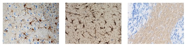 |
| (Left) IHC staining of purified anti-CX3CR1 antibody (clone 8E10.D9) on FFPE normal human brain tissue. IHC staining of purified anti-GFAP antibody (middle; clone SMI 24) or purified anti-Myelin CNPase antibody (right; clone SMI 91) on FFPE mouse brain tissue. Ultra-Streptavidin (USA) HRP kit (Multi-Species, DAB) was used for detection followed by hematoxylin counterstaining |
| Neuroscience Sampler Kits |
|---|
| Glial Cell Marker Antibody Sampler Kit |
| α-Synuclein Antibody Sampler Kit |
| Tau Antibody Sampler Kit |
| P2RY12 is a G-protein-coupled receptor for ADP and ATP that inhibits the adenylyl cyclase second messenger system. Required for normal platelet aggregation and blood coagulation, P2RY12 is also a highly selective marker for microglia that specifically distinguishes these cells from other myeloid cells. |
 |
 |
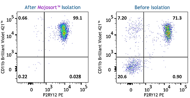
| A single cell suspension from adult C57BL/6 brain was prepared using Trypsin digestion and 70/37/30% percoll gradient for positive selection of P2RY12+ cells using the MojoSort™ Mouse P2RY12 Selection Kit. Cells were stained with either PE anti-mouse P2RY12 and BV421™ anti-mouse CD11b (left, middle). (Right) IHC staining of purified anti-P2RY12 antibody (pink; clone S16007D) on FFPE mouse brain tissue. Nuclei were counterstained with DAPI (blue). |
| P2RY12 Reagents |
|---|
| Human P2RY12 Antibodies |
| Mouse P2RY12 Antibodies |
| MojoSort™ Mouse P2RY12 Selection Kit |
| Additional Reagents |
|---|
| LEGENDplex™ Human Macrophage/Microglia Panel |
| gC1q-R (p33) |
| C3AR |
| C3a/C3a(desArg)/C3 |
| C3/C3b/iC3b/C3d |
| CD11b |
| CD45 |
| CX3CR1 |
 |
 |
 |
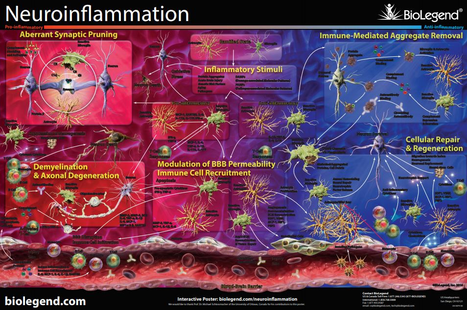
| If you wanted more information on neuroinflammation, check out our intricate poster, which features synaptic pruning. We also have a webpage highlighting the role of the blood brain barrier, neuronal injury, and immune cells on neuroinflammation. Request the poster or visit the webpage for more! |
 |
*Any references to promotions on this page may not be valid at this time. View our promotions page for the most up-to-date promotions.


Follow Us